Download
1 / 42
420 likes | 479 Vues
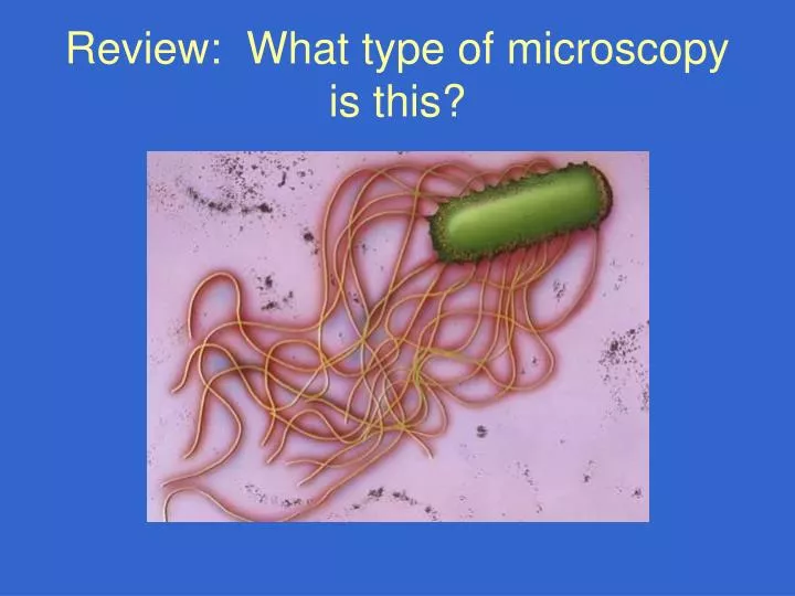
Review: What type of microscopy is this?. Review: What type of microscopy is this?. Review: What type of microscopy is this?. Review: What type of microscopy is this?. Review: What type of bacteria is this?. Review: What type of bacteria is this?. Culture Methods. Culturing Microbes.

E N D
Culturing Microbes • Detection by culture or infectivity assays is preferred • demonstrates that the target microbes are alive and capable of multiplication or replication. From a public health and risk assessment standpoint, microbial pathogen assays based on infectious units are the most relevant and interpretable ones
Culturing Bacterial Pathogens • Continued interest and use because of newly recognized, newly appreciated and evolving agents • Ability to culture some bacterial pathogens goes back more than a century • Salmonella and Vibrio species • Culturing bacterial pathogens from water, wastewater and biosolids remains technologically underdeveloped • Has not advanced greatly beyond the application of methods used in clinical diagnostic and/or food bacteriology • Greater need for more rapid culture methods • Combine culture with molecular detection
Culturing Bacteria Pathogens • Salmonella, Shigella, Campylobacters and Vibrios: culture methods little changed beyond efforts to improve recoveries using modified pre-enrichment and enrichment broths and differential and selective agars • For some other bacterial pathogens: e.g., enterohemorrhagic strains of Escherichia coli (O157:H7): culturing from water a challenge; many other, non-pathogenic strains of E. coli. • select for their growth based on distinctive biochemical or other properties to facilitate separation from the other, non-target strains • e.g., sorbitol-MacConkey Agar for E. coli O157:H7 • Most E.coli ferment D-sorbitol; most E. coli O157:H7 do not; • Sorbitol-negative E. coli O157:H7 colonies are colorless • Non-O157:H7 colonies are pink.
Culture or Infectivity Assays for Bacteria Traditional approach • pre-enrich and/or enrich using non-selective and then selective broth media, or • grow colonies on membrane filters • Then: • Transfer to differential and selective agars • Recover presumptive positive colonies • Biochemical, metabolic and other physiological testing • Serological or other immunochemical typing and identification (agglutination, enzyme immunoassay, etc.) • Other characterization: phage typing, nucleic acid analyses, virulence tests (cell cultures, animal ileal loops, animal infection, etc.)
Enrichment Cultures • Observe for growth by turbidity, clearing, gas production, color change, etc. • Score as presence-absence (positive or negative) • Quantify using replicate and different volumes to compute a Most Probable Number • Example: • 5 x 10 ml • 5 x 1 ml • 5 x 0.1 ml Left: negative Right: positive (color change)
Bacterial Culture • Often 2 step • Presumption • Confirmation • All in one test • Chromogenic media • Enzymatic test (e.g. Β-glucoronidase activity on MUG)
Basic Bacterial Techniques • Streak Plate • For isolation of single colony • Spread Plate • Pour Plate • MPN liquid assay
Problems in Culture Methods for Bacterial Pathogens • Inefficient growth (low plating efficiency), • Slow growth rates • Overgrowth by other non-target bacteria. Efforts to improve culture and reduce or eliminate non-target bacteria: • antibiotics • physical (heat) treatments • chemical (acid) treatments • specialized plating: e.g., dual media plating
Problems in Culturing Bacterial Pathogens Inability of typical culture methods now in use to detect or distinguish: • pathogenic from non-pathogenic strains • sources of pathogens (“microbial source tracking”) • newly emerging pathogenic strains • evolutionary processes and mechanisms • the role of environmental change in selection for or emergence of new pathogenic strains • Antibiotic and disinfectant resistant strains
Pathogenic Bacteria For Which Culture Methods Are Underdeveloped for Food and Water • Pathogenic E. coli • Campylobacter jejuni; other Campylobacters • Yersinia enterocolytica, • Aeromonas hydrophila, • Helicobacter pylori, • Legionella species • Mycobacteriumavium-intracellulare • Shigella Better developed: • Salmonella species • Some pathogenic E. coli O157:H7
Detection of Stressed, Injured and Viable-But-Nonculturable (VBNC) Bacteria • Waterborne bacterial pathogens and indicators are often physiologically altered/stressed and not efficiently cultured using standard selective and differential media • Heavy metals, disinfectants, pH extremes, other environmental stressors • Causes considerable underestimation of the concentrations of these bacteria in water and therefore, underestimation of their risks to human health • Stressed, injured and VBNC bacteria may still be fully infectious for humans and other animal hosts (there is disagreement on this point) • Repair and resuscitation methods improve the detection of viable and potentially cultural bacteria, but, these methods are rarely used to detect pathogens in drinking water.
Assay Methods for Viruses • Electron Microscopy (EM) and Immune EM • Insensitive (>1,000,000 particles/ml) • OK for clinical but not environmental virology • Animal Infectivity • Slow, cumbersome, expensive, ethical considerations • Culture or infectivity • Now widely used in environmental virology • Cytopathogenic effects • Growth, but no cytopathogenic effects • detect viral antigens or nucleic acids • Immunoassays • insensitive for direct detection • Amplify viruses in cell cultures • Nucleic acid assays • insensitive for direct detection by hybridization • Amplify in cell culture or in vitro (PCR or RT-PCR)
Quantifying Human Virus Infectivity is a Challenge • Some infect only humans • Some infect certain experimental animals, too • Some infect experimental animals and cell cultures • Different ratios of infectivity to virions (particles) • 1 infectious unit ~ 1 virus particles • some bacteriophages) • 1 infectious unit ~100 virions: • some cell culture adapted viruses • 1 infectious unit ~10,000-100,000 virions • many “wild-type or field viruses
Detection Of Viral Pathogens by Culture • Viruses are obligate intracellular parasites • Some viruses can be propagated or cultured in susceptible hosts • whole animals • mammalian cells grown in culture • Quantify viruses in animals using quantal methods (e.g., Most Probable Number or MPN); in eggs using “pock” assays on egg membranes (smallpox and vaccinia initially enumerated this way) • Virus assays in cell cultures by quantal (e.g., MPN) or enumerative methods (plaque or local lesion assays)
Virus Detection in Cell Culture • Some viruses (some enteroviruses, reoviruses, adenoviruses and astroviruses) propagate in susceptible host cell cultures and produce morphologically distinct cytopathogenic effects (CPE). Virus plaques in a cell layer overlaid with agar medium and stained with a vital dye Infected Cell Culture with CPE Uninfected Cell Culture
Challenges in Assaying Viral Infectivity • Host susceptibility and variability in host susceptibility • Type of host cells and cell culture assay methods: • Plaque (enumerative) assays • Quantal (liquid culture) assays • Quantal assay endpoints: • Cytopathogenic effects: visible changes in cells • Immunodetection or nucleic acid detection • Animal bioassays: used if no cell culture assay available • Virus titer often increases with serial passages in hosts
Estimating Viral Dose:Relationship of Infectivity to Virus Particle Count • As little a one or a few intact, functional virus particles are capable of causing infection in a susceptible host. • Ratios of virus particles to infectious units are highly variable and are subject to change: • Passage of viruses in susceptible host cells reduces the ratio of virus particles to infectious units • rotavirus: • initial ratio: ~50,000 virus particles/infect. unit • after cell culture passage: ~100 particles/infect. Unit • Norwalk Virus appears to be infectious at doses corresponding to as little as a few virus particles.
Challenges in Assaying Viral Infectivity • Some viruses (some enteroviruses, adenoviruses, rotaviruses, astroviruses and hepatitis A virus) grow poorly or slowly in cell cultures and produce little or no CPE. • Greater detection with additional analytical techniques: • Viral antigens • Immunofluorescence assays, enzyme immunoassays, radioimmunoassays, etc. • Viral nucleic acid assays: hybridization and/or amplification • Combined cell culture + RT-PCR demonstrates presence of greater numbers of infectious viruses than CPE alone • Post-disinfection, more viruses are detected than by CPE
Detection of Hepatitis A Virus in Cell Culture by Radioimmunofocus Assay
Detection of Protozoan Parasites by Culture Environmental forms of some protozoan parasites, such as spores and oocysts, are culturable in susceptible host cells • Culture Cryptosporidium parvum oocysts and some microsporidia spores in mammalian cell cultures; observe living stages • Culture free-living amoebas (Naegleria spp. and Acanthamoeba spp.) on lawns of host bacteria, such as E. coli, on nonnutrient agar; they form local lesions. • For other waterborne parasites, such as Giardia lamblia and Cyclospora cayatenensis, culture from the environmental stage (the cyst or oocyst) is still not possible
Detection of Protozoan Parasites by Culture • Spores of some microsporidia (Encephalitozoon intestinalis) and the oocysts of Cryptosporidium parvum can be cultured in mammalian host cells where spores germinate or oocysts excyst and active stages of the organisms can proliferate. • Living stages detected (after immunofluorescent or other staining) and quantified: score positive and negative microscope fields or cell areas (slide wells), or count numbers of foci of living stages or discrete living stages. • Express concentrations MPNs or other units, such as numbers of live stages. • Detection of living stages also possible by FISH, PCR or immunoblotting • Facilitates molecular characterization
C. parvum Oocysts Immunofocus of Living Stages: in MDCK Cells with C3C3-FITC Antibody
Pathogen Detection by Biochemical Methods • Enzymatic activities unique to target microbe • Signature Biolipid Analysis: • Detection of unique biolipids by gas-chromatography, mass spectrometry and other advanced organic analytical methods • Extract and purify from cells • Analyze • Other biochemical markers unique to a specific pathogen or class of pathogens.
Examples of Other Biochemical Techniques • BIOLOG (carbon/nitrogen/sulfurutilization profiles) • API Biostrips • VITEK and VITEK II • Enzyme activity assays • Colormetric or Fluorescence reporting • FAME-fatty acid methyl ester • FTIR spectroscopy
General Biochemical Targets • Adenylate Kinase/ATP Luminescence • Headspace pressure/gas analysis • CO2 production • Electrical conductance/impedance • UV 260/280 • Excites amino acids with conjugated double bonds (e.g. trytophan, tyrosine, phenylalanine) • Longer wavelengths (Nicotinamides/Riboflavins) • NAD(P) 325-354 excitation; ~480 peak fluorescence • Riboflavin 380-400 excitation; ~520 peak fluorescence
Light Adsorption/Fluorescence Examples of Stand-off/DTW Systems: • BAWS system (UV/visible absorbance and Fluorescence) • FLAPS system (near UV instead of Far to improve signal to noise ratio • LIDAR and UV-LIF • UVRR or Raman Resonance • Raman Spectroscopy • LIBS
Terminology • Biosensor • ACPLA