Download
1 / 37
1.19k likes | 2.65k Vues
TUMORS AND TUMOR-LIKE LESIONS OF INFANCY AND CHILDHOOD . Malignant neoplasms are the second most common cause of death in children between the ages of 4 and 14 years inUSA only accidents exact a higher toll. Benign tumors are even more common than are cancers. . Benign Tumors .

E N D
Malignant neoplasms are the second most common cause of death in children between the ages of 4 and 14 years inUSA • only accidents exact a higher toll. • Benign tumors are even more common than are cancers.
Benign Tumors • any tumor may be encountered in the pediatric age group • occur commonly in childhood are: • Hemangiomas • Lymphangiomas • sacrococcygealteratomas
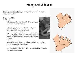
Hemangiomas • are the most common tumors of infancy. • Type: cavernous and capillary hemangiomas • In children most hemangiomas are located in the skin, particularly on the face and scalp • Appear as flat to elevated, irregular, red-blue masses; the flat, larger lesions are referred to as port wine stains. • The vast majority of superficial hemangiomas have no more than a cosmetic significance
Congenital capillary hemangioma at birth at 2 years of age after the lesion had undergone spontaneous regression
Hemangiomas • Rarely, they may be the manifestation of a hereditary disorder associated with disease within internal organs, such as the von Hippel-Lindau and Sturge-Weber syndromes
Lymphangiomas • Represent the lymphatic counterpart of hemangiomas. • They are characterized by cystic and cavernous spaces lined by endothelial cells and surrounded by lymphoid aggregates • They may occur on the skin but, more importantly, are also encountered in the deeper regions of the neck, axilla, mediastinum, and retroperitoneum. • Though histologically benign, they tend to increase in size after birth and may encroach on mediastinal structures or nerve trunks in axilla.
Sacrococcygealteratomas • are the most common germ cell tumors of childhood, accounting for 40% or more of cases • approximately 10% of sacrococcygealteratomas are associated with congenital anomalies, primarily defects of the hindgut and cloacal region and other midline defects • Approximately 75% of these tumors are histologically mature with a benign course, and about 12% are unmistakably malignant and lethal • Most of the benign teratomas are encountered in younger infants (<4 months), whereas children with malignant lesions tend to be somewhat older.
Malignant Tumors • The organ systems involved in infancy and childhood: • the hematopoietic system • neural tissue • soft tissues (common adults tumors: lung, heart, prostate, and colon)
Biological differences between malignant tumors of childhood from those in adults • Relatively frequent demonstration of a close relationship between abnormal development (teratogenesis) and tumor induction (oncogenesis) • Prevalence of constitutional genetic abnormalities or syndromes that predispose to cancer • Tendency of fetal and neonatal malignancies to spontaneously regress • Improved survival or cure of many childhood tumors
Histological differences between malignant tumors of childhood from those in adults • Many malignant pediatric neoplasms tend to have a primitive (embryonal) rather than pleomorphic-anaplastic • Because of their primitive histologic appearance, many childhood tumors have been collectively referred to as small, round, blue cell tumors, include: • neuroblastoma • lymphoma • Rhabdomyosarcoma • Ewing sarcoma (peripheral neuroectodermal tumor) • Wilms' tumor. • There are usually sufficient distinctive features to render a definitive diagnosis on the basis of histologic examination alone, but when necessary, clinical and radiographic findings, combined with ancillary studies (e.g., chromosome analysis, immunoperoxidase stains, and electron microscopy) are used.
Malignant Tumors Neuroblastoma Retinoblastoma Wilms' tumor
Neuroblastoma • Neuroblastomas and related tumors (ganglioneuroblastomas and ganglioneuromas) arise from neural crest-derived cells in the sympathetic ganglia and adrenal medulla. • Neuroblastomas are undifferentiated neoplasms, whereas ganglioneuroblastomas and ganglioneuromas demonstrate evidence of differentiation (Schwannianstroma and ganglion cells). • Neuroblastomas secrete catecholamines, whose metabolites (VMA/HVA) can be used for screening patients.
Neuroblastoma • Neuroblastoma is composed of small cells embedded in a finely fibrillar matrix (neuropil). with Homer-Wright pseudo-rosette. • Ganglioneuromas, arising from spontaneous or therapy-induced maturation of neuroblastomas, are characterized by clusters of large cells with vesicular nuclei and abundant eosinophilic cytoplasm (arrow), representing neoplastic ganglion cells. Spindle-shaped Schwann cells are present in the background stroma.
Neuroblastoma • Age and stage are the most important prognostic features; infants usually have a better prognosis than older children, while children with higher stage tumors fare worse.
Neuroblastoma • Children younger than 2 years with neuroblastomas generally present with protuberant abdomen resulting from an abdominal mass, fever, and weight loss. • In older children the neuroblastomas may remain unnoticed until metastases cause hepatomegaly, ascites, and bone pain.
Retinoblastoma • is the most common malignant eye tumor of childhood. • Retinoblastoma frequently occurs as a congenital tumor • it can be multifocal and bilateral • it undergoes spontaneous regression • patients have a high incidence of second primary tumors. • The incidence decreases with age, most cases being diagnosed before the age of 4 years.
Retinoblastoma • Occur in both familial and sporadic patterns • Familial cases • develop multiple tumorsthat are bilateral, although they may be unifocal and unilateral. • increased risk for developing osteosarcoma and other soft tissue tumors Sporadic cases All of the sporadic nonheritable tumors are unilateral and unifocal.
Retinoblastoma • Approximately 60% to 70% of the tumors are associated with a germline mutation in the RB1 gene and are hence heritable. The remaining 30% to 40% of the tumors develop sporadically, and these have somatic RB1 gene mutations.
Retinoblastoma cohesive tumor in retina abutting optic n High power view showing Flexner-Wintersteiner rosettes (arrows) and numerous mitotic figures.
RetinoblastomaClinical features • The median age at presentation is 2 years, although the tumor may be present at birth. • The presenting findings include poor vision, strabismus, a whitish hue to the pupil and pain and tenderness in the eye. • Untreated, the tumors are usually fatal, but after early treatment with enucleation, chemotherapy, and radiotherapy, survival is the rule. • Some tumors may spontaneously regress • patients with familial retinoblastoma are at increased risk for developing osteosarcoma and other soft tissue tumors.
Wilms' tumor • Wilms' tumor, or nephroblastoma, is the most common primary tumor of the kidney in children. • Most cases occur in children between 2 and 5 years of age. • This tumor illustrates several important concepts of childhood tumors: • the relationship between congenital malformation and increased risk of tumors • the histologic similarity between tumor and developing organ • the remarkable success in the treatment of childhood tumors
Wilms' tumor • Three groups of congenital malformations are associated with an increased risk of developing Wilms' tumor: • Patients with the WAGR syndrome: aniridia, genital abnormalities, and mental retardation • Denys-Drash syndrome (DDS): gonadaldysgenesis and renal abnormalities • Beckwith-Wiedemann syndrome (BWS) enlargement of individual body organs (e.g., tongue, kidneys, or liver) or entire body segments (hemihypertrophy); enlargement of adrenal cortical cells (adrenal cytomegaly)
Wilms' tumor • WAGR and DDS are associated with WT1 inactivation, while BWS arises through imprinting abnormalities at the WT2 locus.
Wilms' tumor in the lower pole of the kidney Tumor shows the characteristic tan to gray color and well-circumscribed margins
Triphasic histology of Wilms' tumor: • the stromal component is composed of spindle-shaped cells • the immature tubule of the epithelial component • the blastema : the tightly packed blue cells Nephrogenic rests are precursor lesions of Wilms' tumors
Wilms' tumor • palpable abdominal mass • fever • abdominal pain • hematuria
Prognosis of Wilms' tumor • is generally very good, and excellent results are obtained with a combination of nephrectomy and chemotherapy. • Diffuseanaplasia is a harbinger of adverse prognosis