Download
1 / 68
680 likes | 810 Vues
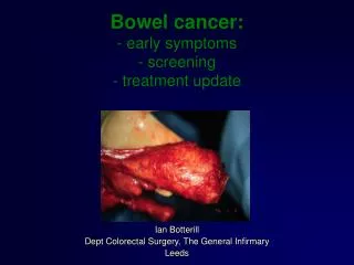
Pathology Issues in Bowel Screening. Frank Carey. SPAN. Lead-time Bias. Tumour Growth. Time. Proving Screening Works. Population-based randomised trials in which the whole group offered screening (including refusers and interval cancers) is compared with the control group.

E N D
Pathology Issues in Bowel Screening Frank Carey SPAN
Lead-time Bias Tumour Growth Time
Proving Screening Works Population-based randomised trials in which the whole group offered screening (including refusers and interval cancers) is compared with the control group
Disease-Specific Mortality in gFOBT Randomised Trials (Relative Risks) • Minnesota • Annual 0.67 (CI 0.51-0.83) • Biennial 0.79 (CI 0.62 - 0.97) • Nottingham • Biennial 0.85 (CI 0.74 - 0.98) • Funen • Biennial 0.82 (CI 0.68 - 0.99) • Göteborg • Biennial 0.84 (CI 0.71-0.99)
A Scottish Tradition…..Robert Burns “Death and Dr Hornbrook” (1785) "Ev'n them he canna get attended, Altho' their face he ne'er had kent it, Just shite in a kail-blade, an' sent it, As soon's he smells 't, Baith their disease, and what will mend it, At once he tells 't.
Scottish Pilot 2000-07 Fife, Grampian, Tayside FOB kits posted out and returned to Dundee +ve tests lead to colonoscopy performed locally
Rate ratio of Colorectal Cancer invited vs controls Overall 0.90 (0.830 – 0.989) Relative reduction in CRC mortality 10% Participants only 0.73 (0.653 – 0.824) Relative reduction in CRC mortality 27%
Organisation of the bowel cancer screening programme - Scotland Single Centre Investigation and treatment devolved to health boards (n=14) Age range 50 - 74
Make a diagnosis Plan treatment and follow up Collect accurate data Audit of service development Facilitate high quality research Pathology
Quality Measures in Bowel Screening • Key Performance Indicators (KPI) • High level overview of programme performance • Endoscopy (“JAG” accreditation) • Pathology
Key Performance Indicators (KPIs) Uptake overall by deprivation category response rate to first invitation response rate to reminders Time to colonoscopy Proportion of +ves undergoing colonoscopy Colonoscopy completion rate Colonoscopy complication rate admissions perforations bleeding deaths Positivity rate Cancer Detection Rate Stage at diagnosis (incl. polyp cancers) Adenoma detection rate overall high risk PPV for cancer for adenoma for high risk adenoma for any neoplasia
KPI 4 Positive screening test result rate, by NHS Board
Pathology QA • Adherence to RCPath standards in reporting of colorectal cancers • Participation in web-based EQA • Central referral of cases suspected/diagnosed as polypoid cancer (T1Nx) Close links with other UK jurisdictions
EQA An essential part of quality assurance for the programme All UK countries participate (+ Irish Republic, Slovenia) Our first experience of electronic (web based) EQA Administered by Dr Nic Mapstone, hosted by University of Leeds
EQA Slides accessed online http://www.gieqa.org.uk/ 4 possible answers for each slide Low grade dysplasia High grade dysplasia Adenocarcinoma Other It is possible to enter a comment
EQA A case is valid only if the diagnosis is agreed by 80% of the regional lead pathologists Scores per case: 2 points for same diagnosis as consensus 1 point for one category removed (e.g. high grade dysplasia/carcinoma) 0 points otherwise
Case E8 Result
Scottish participation in EQA • 43 registered • Limited data on actual participation (July 2012 review of circulations G,H) • 1/3 participated in both • 1/3 in one of the two circulations • 1/3 in neither • Updated data awaited.
Slide referral • Recognised difficulty in distinguishing epithelial misplacement from invasive cancer in adenomatous polyps
Case D9 Result
Referral Panel • Dr M Balsitis • Prof F Carey • Dr P Fineron • Prof G Murray • (Dr A Lessels)
Referral review • Started April 2009. 240 cases received by March 2012 • Participation not even across Boards.
30 cases (12.5% of total) submitted with a favoured diagnosis of cancer were assessed as benign by the panel • 10 cases (4.2%) submitted as probably benign were upgraded to cancer • There was disagreement among panel members as to the final diagnosis in 22 cases (9.2%). All cases were seen in the first instance by 2 pathologists. In the cases where discrepant diagnoses were made a third panel member was involved and the majority diagnosis was registered as final. • 4 cases (1.6%) were referred to the English/Welsh Expert Panel. These were difficult, complex cases. 3 were finally diagnosed as benign and one as cancer.
Case F7 • Result
Adenomas in Screening • Adenomas much more common than cancers • Adenomas are the precursors of most cancers • Adenomas (even when removed) are a marker of cancer risk • The programme is almost as much about adenomas as cancer
Issues in adenomas Recognising adenomas Categorising adenomas Serrated lesions
Risk of Advanced Neoplasia at5.5 yrs in a Colonoscopic Series Finding at first exam Patients Ad Neo RR No neoplasia 298 7 1 Tubular Adenoma <10mm 622 38 2.56 1-2 496 23 1.92 3+ 126 15 5.01 Tubular Adenoma >10mm 123 19 6.40 Villous Adenoma 81 13 6.05 High Grade Dysplasia 46 8 6.87 Carcinoma 23 8 13.56 Lieberman et al 2007
Grading Dysplasia in 2189 Adenomas at 13 Centres min max median mild 29% 88% 42% moderate 10% 67% 43% severe 1% 24% 4%
Histology of 2206 Adenomas at 13 Centres min max median tubular 62% 93% 84% tubulovillous 6% 37% 15% villous 0% 6% 1%
Tubulovillous Adenomas The 20% Rule (for intact excised lesions): The minor component comprises at least 20% of the lesion.
“Advanced” Adenoma Patients > 1 cm (measured for smaller lesions on microscope slide) multiple polyps villous component* severe dysplasia* *in selected series only
OPTICAL PROJECTION TOMOGRAPHY Original prototype was invented by James Sharpe whilst at MRC Human Genetics in 20021 Whole mount, in vitro, imaging technology for small biological specimens (1-15mm) Optical equivalent of an X-ray CT scanner Generates 3D images and 2D virtual sections through three orthogonal planes Visualises unstained tissue as well as fluorescent labels (emission mode) and coloured stains (transmission mode). Ideal for analysis of gene and protein expression. 1Sharpe et al 2002 296, 541-545
The Imaging Gap 10 μm 100 μm 1mm 1cm 10 cm OPT CONFOCAL MICROSCOPY MRI/CT ORGANS EMBRYOS ORGANISMS TISSUES CELLS
FEATURES OF OPT Produces 3D images & virtual sections in 3 orthogonal planes Wholemount technology Non-destructive – analysis post OPT possible (e.g. IHC) Visualise unstained anatomy with autofluorescence* Visualise fluorescent labels & coloured stains Investigate gene & protein expression in context of 3D anatomy Produces quantifiable and digital data – archive Digital images to scroll through, send for opinions or as teaching tools *Unstained sections used for the purposes of this study. 1Sharpe et al 2002 296, 541-545
How does OPT work Two Imaging Modes: Transmission i.e.Brightfield Emission i.e.Fluorescent
How does OPT work Two Imaging Modes: Transmission i.e.Brightfield Emission i.e.Fluorescent