Download
1 / 29
290 likes | 711 Vues
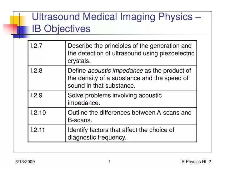
Ultrasound Medical Imaging Physics – IB Objectives. Ultrasound Production and Detection. Based on piezoelectric effect. From http://en.wikipedia.org/wiki/Medical_ultrasonography. Piezoelectric Effect in Crystals. Applied electric field produces mechanical vibration

E N D
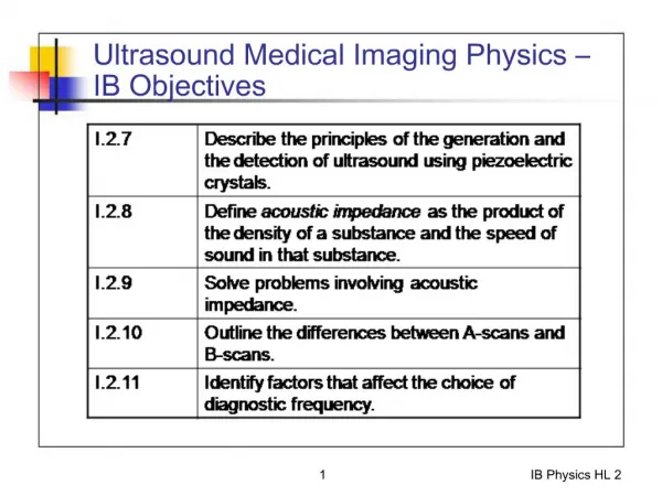
Ultrasound Medical Imaging Physics –IB Objectives IB Physics HL 2
Ultrasound Productionand Detection • Based on piezoelectric effect From http://en.wikipedia.org/wiki/Medical_ultrasonography IB Physics HL 2
Piezoelectric Effect in Crystals • Applied electric field produces mechanical vibration • Also, mechanical vibration produces electrical signal • Single crystal can be both ultrasound source and detector • Not at exactly same time • Mechanical vibration moves at same frequency as electrical vibration (1 MHz to 20 MHz) + + + + + + Piezoelectric crystal (e.g., quartz) - - - - - - - Electrodes IB Physics HL 2
Wave Motion in Solids • After piezoelectric crystal starts sound wave (ultrasound wave?), wave travels through tissue • Wave may reflect, refract, or be transmitted between two different materials (organs, tissue types, etc.) • Acoustic impedance (~index of refraction) • Acoustic impedance (Z) is product of Density of medium and Speed of wave:Z = v [units of kg m-2 s-1] [Rayl] • Ex: waterSpeed is 1,480 m/s; = 998 kg/m3Zwater = 1.48 x 10⁶ kg m-2 s-1 IB Physics HL 2
Reflection and Transmission ofWaves with Ultrasound • Reflection and transmission:When wave goes from medium with impedance Z1 to a medium with impedance Z2 • Reflection fraction: (Z2 – Z1)2/(Z2+Z1)2 • Transmission fraction: (2Z2)2 / (Z2 + Z1)2 • Limiting cases: • If Z1 = Z2, no reflection, and transmission = 1 • Reflection fraction + transmission fraction = 1 • Note: acoustic impedance is frequency-dependent IB Physics HL 2
Reflection and Transmission ofWaves with Ultrasound - Examples • What is fraction of sound reflected and transmitted when • Sound travels from water to muscle (Z muscle = 1.7 x 10⁶ kg m-2 s-1) • Sound travels from water to air (Z air = 400 kg m-2 s-1) • Note: acoustic impedance is frequency-dependent IB Physics HL 2
Scan Modes with Ultrasound • A Mode (Amplitude modulation) • B Mode (Brightness mode) • M Mode (Moving mode) • Doppler (Doppler imaging) IB Physics HL 2
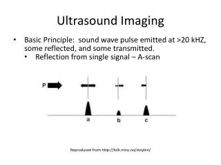
Scan Modes with Ultrasound • A Mode: Amplitude modulation • Single transducer generates ultrasound, receives ultrasound • Information is available in only one-dimensional scan Height of returning pulseproportional to strengthof returning pulse IB Physics HL 2
Scan Modes with Ultrasound • A Mode: Amplitude modulation • Assuming speed of sound in muscle / soft tissue is 1,540 m/s • How far under the skin does the organ start? • How long is the organ? 35 s 0.18 ms IB Physics HL 2
Scan Modes with Ultrasound • B Mode (Brightness mode) • Several transducers on handgrip record travel time simultaneously • Can build up 2-D picture of reflections • Brightness of image on screen is proportional to strength of reflection Transducers IB Physics HL 2
Scan Modes with Ultrasound • B Mode (Brightness mode) • Several transducers on handgrip record travel time simultaneously • Can build up 2-D picture of reflections IB Physics HL 2
Ultrasound Frequency Choice • High frequency - high resolution • Get more detail with a higher frequency scan than a lower frequency scan • High frequency – high attenuation • Higher frequencies are attenuated faster than lower frequencies • Get more penetrating images using lower frequencies IB Physics HL 2
Ultrasound Imaging - Cautions • Difficult to get imaging from brain • High-intensity scans can transfer energy to object being scanned • Potential warming / damage to imaged object • Fetus IB Physics HL 2
Ultrasound Imaging – Other uses • Doppler shift scans • Determine blood flow speed • High speed – indication of blockage • Moving ultrasound (M Scan) • Real-time image of moving objects • E.g., heart beating • Fetus IB Physics HL 2
Ultrasound - Key Ideas IB Physics HL 2
Magnetic Resonance Imaging (MRI) • Also called Nuclear Magnetic Resonance (NMR) scan IB Physics HL 2
NMR Scans –IB Objectives IB Physics HL 2
Fundamental Concept - Magnetic Energy • Atoms in imaged object, especially hydrogen atoms, have a magnetic moment (~a compass) • Magnetic moment is a consequence of the spin of the proton • No classical analog • Charge in motion produces magnetic field • Like a compass, the magnetic moments of the hydrogen atoms want to line up in the direction of the applied magnetic field • The stronger the field, the more the atoms line up with it IB Physics HL 2
N N N N N Fundamental Concept - Magnetic Energy Appliedmagnetic field Most of the atoms in the sampleare oriented in the direction ofthe magnetic field IB Physics HL 2
Appliedmagnetic field N N N N N Fundamental Concept - Magnetic Energy When atoms shift theirmagnetic fields to beopposite the external field,they gain energy (photon). When atoms shift their magneticfields to be along the externalfield, they give up energy (photon). IB Physics HL 2
MRI / NMR Scanner • NMR scanners send in a radio signal in to the sample, with just the right amount of energy to flip the nuclear magnetic moments back an forth, from opposite to along the magnetic field. • Resonance effect • Frequency is called the Larmor frequency • Able to localize the resonant area with slightly deformed magnetic fields • Gradient fields IB Physics HL 2
MRI Scanner - Operation • Scanner detects where large numbers of hydrogen atoms are • ~Water • Builds up 2-D image of object / body, which can be converted into a 3-D image • Resonance of hydrogen nuclei also sensitive to nearby atoms (electrons) • Distinguish compounds that hydrogen is in IB Physics HL 2
MRI Scanner - Details • Useful for imaging skull and brainWhole-body diagnosis IB Physics HL 2
MRI Simulation - Questions • What is the relationship between the applied external magnetic field, and the frequency of the radio-wave energy that flips the spins? • Direct, inverse, or no relation • How do the fringe fields help localize the RF signal from the body? • TUMOR HUNT: • Uncheck “Show atomic nuclei” • Click “Add tumor” • Look for evidence of tumor in RF signal IB Physics HL 2
MRI Scanner - Cautions • Non-ionizing radiation • Intense magnetic field • No magnetized objects or metal IB Physics HL 2
MRI - Key Ideas IB Physics HL 2
MRI - Homework • Write a 1-2 paragraph summary of NMR scans. • Include: • Hydrogen magnetic moment • External magnetic field • Energy of 50 MHz radio photon • Gradient fields (optional) IB Physics HL 2
Scanning Techniques • Excellent table and discussion, p. 502 IB Physics HL 2
Diagnostic and Therapeutic Lasers • Excellent table and discussion, p. 501 IB Physics HL 2