Download
1 / 22
220 likes | 492 Vues
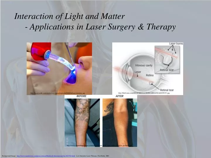
Interaction of Light and Matter - Applications in Laser Surgery & Therapy. http://local.yodle.com/articles/wp-content/uploads/2009/04/laser-dentistry.jpg. http://blstb.msn.com/i/8F/4C8BB16A15B5B2ABEA45EA6685FCF1.jpg.

E N D
Interaction of Light and Matter - Applications in Laser Surgery & Therapy http://local.yodle.com/articles/wp-content/uploads/2009/04/laser-dentistry.jpg http://blstb.msn.com/i/8F/4C8BB16A15B5B2ABEA45EA6685FCF1.jpg http://www.miamiskinandlaser.com/images/before_after_laser_tattoo_removal_treatment.jpg Background Image: http://www.marketwire.com/press-release/Meditech-International-Inc-883754.html: Low Intensity Laser Therapy, Fred Kahn, MD
Outline • Introduction to photomedicine • Understand how light interacts with body tissue and the origins of absorption. • Understand how biological molecules absorb light. • Photocoagulation & Photovaporization • Combine endoscopic and laser techniques to do laser surgery. • Ophthalmological lasers to perform a diabetic retinopathy. • Laser eye surgery – LASIK eye surgery • Lasers in dermatology - tattoo removal • Lasers in dentistry
Introduction to Photomedicine • Photomedicine is the study and subsequent treatment of diseases in the body by exposing the body to light. • In addition this can include diagnostic and therapeutic applications using light for the detection and curing of disease, or phototherapy. • So, if all photons are light, why do some photons produce harmful medical effects in some instances (such as skin cancer), but helpful effects in others (such as vitamin D production)? • Can I control the effects of the light energy in photomedicine? • How do you maximize the effects that seem to be beneficial while minimizing the ones that are harmful? • The answers depends on how light is able to be absorbed by biological tissues and this is due to the properties of the electronic states associated with these biological tissues.
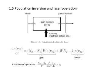
Interaction of Light and Matter • Why do I care how light and matter interact? • Laser surgery and laser therapy involve the transfer of energy from the light to the body’s tissues through several mechanisms. • There are five effects that can happen to light when it interacts with matter. • - reflection off of a material. • - transmission through a material. • - absorption by the material. • - scattering by the material. • - re-emission by the material. http://www.photobiology.info/Photomed_files/Fig4.png
Interaction of Light and Matter • We are concerned with the scattering, absorption, and re-emission of light as the light travels through tissues. • - Photons of light can be absorbed by electrons in the tissue material. This takes photons out of the beam of transmitted light and the intensity of the beam diminishes. • - Absorbed photons can be re-emitted and these re-emitted photons can be in any direction. This also contributes to the lowering of the transmitted beam intensity. • - Photons do not have to get absorbed (and eventually re-emitted) by electrons in the tissue material. Instead they can be scattered off of electrons (like pool balls) and if photons are scattered away from the transmitted beam, the beam intensity decreases. • Here we are concerned with the intensity of light and further we’ll be interested in the transfer of photon energy to body tissue.
Interaction of Light and Matter - Light Intensity • Recall that the intensity of light is defined as the power per unit area of delivery, or the energy that is delivered to a spot of area A in a time t. • Imagine that you turn on the incandescent* lamp in your room and that you stand at arms length from the light. Suppose further that your bulb is rated at 100 W (so that it is really bright and keeps you up at night.) What is the intensity of the light that passes out through a sphere centered on the light bulb at a distance of say 1m? • Now compare this to a milliwatt laser at the same distance away. How many times more/less intense is the laser beam compared to the light from the bulb? * (Does anyone still have these anymore?)
Interaction of Light and Matter - Light Intensity • Let’s do one more calculation. • Suppose that you are looking at the sun with your eyes (or worse yet using binoculars, a camera lens or a telescope - don’t ever do this by the way) and that you happen to know that the intensity of the sun’s rays at the earth’s surface is 1370-W/m2 (called the solar constant.) Further your eye is a convex lens and this lens has a focal length of about 2-cm (the distance from the front of the eye to the retina.) What is the intensity of light on your retina? • How many times more intense is this compared to the light bulb or the laser? • Where does this energy (that’s deposited every second) go? How does it show up in the tissue?
Interaction of Light and Matter - Photocoagulation • When light is incident in body tissue it usually gets deposited in the form of heat and this raises the temperature of the spot and the surrounding tissue where the light is incident. • This heating can cause damage to the proteins in your body especially the myoglobin protein found in muscle. • Proteins are hydrocarbon chains (amino acids) linked together by chemical bonds and if the structure of the protein changes so too does the protein’s function. • Myoglobin has an alpha helix structure which can be viewed as being enclosed in this blue sheath (which is only for illustration). • That helix folds back upon itself into what's referred to as the tertiary structure of myoglobin. • Bonds between the side groups of the amino acid residues (those chains that form the protein) are responsible for holding together the tertiary structure of this protein. http://dl.clackamas.edu/ch106-08/tertiary.htm
Interaction of Light and Matter - Photocoagulation • When exposed to temperatures above about 120oF (60oC) the myoglobin proteins begin to uncoil and the myoglobin loses its structure and the ability to perform its biological function (which is to increase the rate of O2 transfer from the blood into the muscle fibers) . • When muscle tissue is heated the myoglobin denatures (uncoils) and the the structural integrity of the muscle proteins are lost – and the muscle becomes easier to pull apart. • We’ve essentially cooked the muscle tissue with a laser – a process called photocoagulation and the color of the tissue changes. • The cells in the region of tissue hit by the laser beam usually dies and the resulting region of tissue burn is called a photocoagulation burn. • Photocoagulation burns are used to destroy tumors, treat eye conditions and stop bleeding.
Interaction of Light and Matter - Photocoagulatve Trade-offs • In order to effect a local increase in temperature of a region of tissue, energy has to flow into the tissue at a larger rate then the energy leaves that region. • If the relaxation time were really short then heat flows out of the region very fast. • I would like to deliver a high enough dose of energy to a piece (defined by the beam area) of tissue to cause it to coagulate, and this energy (per unit area of beam) is determined by the product of the intensity (power density) and the exposure time. • The surgeon needs to determined how much energy needs to be delivered to the region of interest. • There is a trade off between selecting a high enough intensity (to do the job) while also minimizing the exposure time (to minimize heating effects of the surrounding tissue.)
Interaction of Light and Matter - Photocoagulatve Trade-offs • If the power density is too low, then the exposure time has to be very long to achieve the desired coagulative effects. • A long exposure time will lead to heat flowing into the surrounding tissue leading to damage of a larger area than expected. This can be easily seen. • Conversely, a high power density requires a low exposure time, so the heat flow will be minimized, but the coagulated region may be deeper that required and damage will be deeper. This cannot be easily seen.
Interaction of Light and Matter - Photovaporization – cutting with light • When moderately high intensities ( ~ 10 – 100 W/cm2) are used the tissue exposed to the beam cooks, or photocoagulates. • If extremely high intensities ( > 100 W/cm2) are used we can cause water in the tissue to boil. This is called photovaporization (or photoablation or photodisruption), or the vaporization of water by using a laser beam. • The boiling of water in the tissue turns the tissue into a gas. Thus we can use a laser beam to make a fine incision or to remove layers of tissue cells. • The surgeon has to make sure that there is a small exposure time, but yet that enough energy is delivered to the area to vaporize the cells. • Since we have a high intensity, the heat flows out of the surrounding tissue, albeit not very far, and causes a photocoagulation burn resulting in a bloodless procedure.
Interaction of Light and Matter - Treatment of Diabetic Retinopathy • A common complication of diabetes affecting the blood vessels in the retina (the thin light-sensitive membrane that covers the back of the eye). If untreated, it may lead to blindness. If diagnosed and treated promptly, blindness is usually preventable. • There are two stages of diabetic retinopathy -- nonproliferative and proliferative retinopathy: • Nonproliferativeretinopathy is the earlier stage. In this stage there may be hemorrhages (bleeding) in the retina with leakage of blood causing a "wet retina" or protein deposits (exudates) in the retina. As a consequence, the retina does not receive enough oxygen. This early stage of diabetic retinopathy usually produces no visual symptoms but, if there is fluid in the central portion of the eye (macular edema), vision is diminished. • Proliferative retinopathy is the second stage. New abnormal vessels develop in the retina and grow towards the center of the eye. These vessels frequently bleed into the vitreous (the clear jelly in the center of the eye). Such bleeding episodes cause severe visual problems. Small bleeds may clear up on their own but larger bleeds need surgery. The abnormal vessels may also produce large scars in the retina that may cause the underlying retina to detach (retinal detachment).
Interaction of Light and Matter - Treatment of Diabetic Retinopathy http://www.neec.com/Images/Photos/diabetic_retinopathy_fig_4.jpg http://www.theberries.ca/Archives/2006Winter/images/diabetic_ret1.jpg Nonproliferativeretinopathy Proliferativeretinopathy http://www.neec.com/Images/Photos/diabetic_retinopathy_fig_5.jpg
Interaction of Light and Matter - Treatment of Diabetic Retinopathy • Treatment is by laser surgery. • In the early nonproliferative stage, the laser is used to treat the leaking blood vessels. The laser produces a small scar that helps seal the leak. This is called as focal laser therapy. • In the later proliferative stage, the laser is used to produce many small burns all around the edge of the retina. The aim is to produce disappearance of the new vessels by a procedure panretinal photocoagulation. • Laser therapy can only stop the progression of diabetic retinopathy. It cannot reverse the damage already done. The progression of the retinopathy can be slowed down by careful control of the diabetes, effective reduction of high blood pressure together with regular eye exams and, if needed, prompt laser therapy.
Interaction of Light and Matter - Treatment of Diabetic Retinopathy http://ohiovalleyeye.com/services/eyeinfo_.htm http://www.mountnittany.org/assets/images/krames/139114.jpg The laser light is directed through a special contact lens and directed to the abnormal blood vessels. These vessels are heated and the heating photocoagulates the blood vessels to stop the progression of the disease. http://www.alcon.com/en/img/eye-health/diabetic_retinopathy_t.gif Video session that focuses on the treatment of diseases of the retina and vitreous including macular degeneration, diabetic retinopathy, and retinal degenerations by David Saperstein, M.D., assistant professor, Ophthalmology, University of Washington. http://www.researchchannel.org/asx/uw_ogr_retinal_1300k.asx
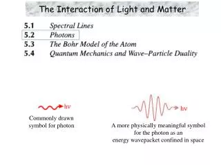
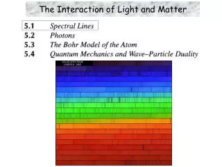
Origins of Absorption • In order for a laser’s light to cause damage, the light has to be absorbed by the tissue. • Laser light is emitted in a specific color – meaning it has a well defined frequency or wavelength. • Most lasers operate from the IR to the UV range in the EM spectrum. • The spectrum on the left is the continuous emission spectrum of an incandescent light bulb and the spectrum on the right is the emission spectrum of a He-Ne laser. • The color of an object is due to the reflection or transmission of different colors of light. For example, a fire truck appears red because it reflects red light and absorbs more green and blue wavelengths. http://academics.hputx.edu/chem/Sample%20Light%20Emission_files/image004.jpg http://images.absoluteastronomy.com/images/encyclopediaimages/h/he/helium_neon_laser_spectrum.png
Origins of Absorption • Another example is your pupil. It appears black because all of the light that falls onto it gets absorbed and transmitted into the eye and almost none gets reflected back. • The wavelengths of light that get absorbed determine the color of an object. • For biological materials their absorption properties depend on wavelength and this gives them their characteristic color and determines which color laser light they will preferentially absorb. • Molecules generally have much more complex electronic structures than single atoms. There electronic structure stabilize the orbitals and generally transitions in molecules from the ground state of the molecule to excited molecular states cost more energy. • This shifts the emission and absorption of light in these molecules to shorter wavelengths.
Origins of Absorption - Selective Absorption in Surgery • Challenge is to find a laser that is capable of operation at an application specific wavelength so that it can be absorbed by body tissue. • Each specific biologic tissue has its own unique absorption properties. • Most of body tissue is water – water is transparent to visible light – it transmits all of it and absorbs very little in the visible portion. • Water however does absorb strongly in the infrared (above 1300nm) and in the ultraviolet (below 300nm). Compare this with the fact that you can see the sun through the atmosphere – the water vapor in the atmosphere doesn’t absorb the visible radiation. • So visible is not so good for absorption by water dominated structures.
Origins of Absorption - Selective Absorption in Surgery • Proteins absorb strongly in the UV. • Blood, dominated by Heme, is an iron complex which absorbs strongly in the visible range. • Body tissue is composed of water and proteins so most body tissues absorb strongly in the UV and in the Blue/green portion of the spectrum and transmit reds and IR. • A surgeon can select a particular laser to target cells (perhaps cancer) for photovaporization by determining which wavelengths your damaged cell will absorb and what the surrounding tissue/cells won’t. • This makes for very precise surgeries and therapeutic techniques.
Origins of Absorption - Selective Absorption in Surgery • Consider as an example the a cross section of the retina. • For ophthalmological surgeries one could use red light (676-nm) from a krypton laser, which is absorbed by melanin. Consequently these layers will be damaged by the red laser light and the surrounding tissue will be unaffected. • Melanin is a brown skin pigment that is produced by cells called melanocytes. Melanin provides some protection against skin damage from the sun, and the melanocytes increase their production of melanin in response to sun exposure. Freckles, by the way, which occur in all peoples, are small, concentrated areas of increased melanin production. • In contrast, the green light of an argon laser (488 & 514-nm)would be absorbed preferentially by blood leading to photocoagulation.
Summary • We’ve looked at photomedicine, or the application of lasers to the treatment of disease. • The intensity of the laser light can be controlled and focused for precise surgical techniques such as laser cutting (photovaporization) and photocoagulation. • These techniques are all due to the interaction of light and matter. • Next we’ll look at using lasers in dermatology, dentistry and eye surgery. • Homework: For Wednesday, September 18, 2012 • Read Chapter 3, sections 3.12 – 3.19 and do • Questions: Q3.1, Q3.2, Q3.3, & Q3.5 • Problems: P3.3 & P3.5