Download
1 / 22
230 likes | 253 Vues
Unit 4: Biological Psychology. Essential Task 4-6 :

E N D
WHS AP Psychology Unit 4: Biological Psychology Essential Task 4-6: Detail historic and contemporary research strategies and technologies that support research (case studies like Phineas Gage, split-brain research, sleep research (EEGs), structural imaging (CAT Scans and MRIs), and functional imaging (PET scans and fMRIs).
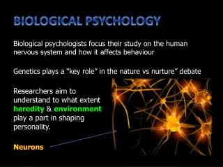
Endocrine System Evolutionary Building Blocks Genetics Biological Psychology Neurons Neurotransmitters Nervous System Central Nervous System Peripheral Nervous System Motor Sensory Spinal Cord Brain Autonomic Somatic We are here Brain Imaging Sympathetic Parasympathetic
Essential Task 4-6: Outline • Detail historic case studies like Phineas Gage and split-brain research • Contemporary research strategies and technologies • lesions • microelectrodes • sleep research (EEGs) • structural imaging (CAT Scans and MRIs) • functional imaging (PET scans and fMRIs)
Case Studies: Phineas Gage Outline
Gage Outline
Split Brain Research Outline
Lesion Outline Techniques to Study the Brain A brain lesion experimentally destroys brain tissue to study animal behaviors after such destruction. Hubel (1990)
Microelectrode Techniques Outline • Very small electrodes inserted into individual neurons • Used to study activity of a single neuron
EEG (Electroencephalogram) Outline • Macroelectrode Techniques • Used to get a picture of overall activity in the brain • An example is an which uses electrodes placed on a person’s scalp to measure an amplified recording of the electrical waves sweeping across the brain’s surface.
Sleep Research Outline
Sleep Research Outline
EEG imaging Outline • 21 Sensors on the scalp record changes in electrical activity and feed them into a computer. The computer translates them into color and motion on a map of the brain displayed on a television monitor
Brain Imaging Outline Structural Imaging Functional Imaging CAT Scan MRI PET Scan fMRI
CAT Scans Outline • Computerized Axial Tomography (CAT-scan) • Uses X-rays to create a 3-dimensional image of the brain • CT scans can often show the size and locations of brain abnormalities caused by tumors, blood vessel defects, blood clots, strokes and other problems.
More CAT Scans Outline
Not a CAT Scan Outline
MRI – Magnetic Resonance Imaging Outline • Magnetic Resonance Imaging (MRI) • Uses a magnetic field and radio waves to produce computer-generated images • They distinguish among different types of brain tissue. Outline
CAT scan vs. MRI CAT scan MRI Outline • Less expensive than MRI • MRI contrast materials used for image enhancement have very low incidence of side effects • Less sensitive to patient movement Give you the structure of the brain • More sensitive to patient movement • CT can be performed if you have an implanted medical device of any kind
PET Scans Outline • Positron Emission Tomography (PET) • Use radioactive glucose to determine location of greatest brain activity PET Scan ofNormal Brain PET Scan of Alzheimer's Disease Brain
fMRIs Outline • Functional Magnetic Resonance Imaging (fMRI) • Shows function and structure by measuring movement of blood molecules within the brain
Anticipation of doing math causes pain in some people. Outline UChicago researchers have found that the higher a person’s anxiety about math, the more anticipating math activated areas of the brain related to experiencing pain. posterior insula -- a fold of tissue located deep inside the brain just above the ear that is associated with registering direct threats to the body as well as the experience of pain.