Download
1 / 31
310 likes | 311 Vues
Explore the discovery of DNA as the genetic material and its structure. Learn about nucleotides, DNA vs. RNA, Chargaff's rules, DNA structure, and organization of genetic material.

E N D
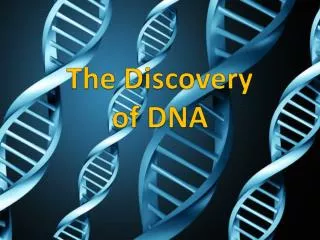
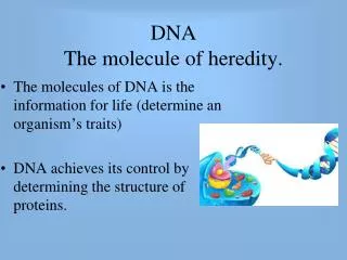
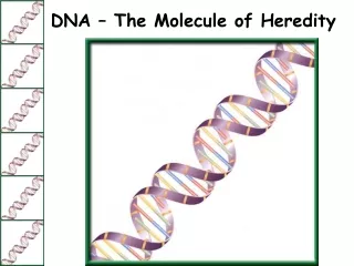
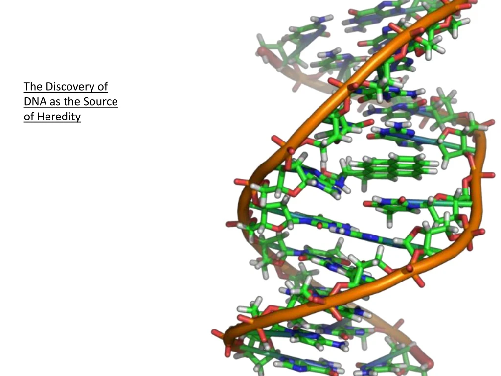
The Discovery of DNA as the Source of Heredity
The human genome project which has decoded the base sequence for the whole 6 feet of the human genome requires a data warehouse (pictured) to store the information electronically. Scientists have programmed nearly 500,000 DVD’s worth of data into 1 gram of DNA!
Confirmation of DNA 1952 | 1969 Hershey • Hershey & Chase • classic “blender” experiment • worked with bacteriophage • viruses that infect bacteria • grew phages in 2 media, radioactively labeled with either • 35S in their proteins • 32P in their DNA • infected bacteria phages Why useSulfurvs.Phosphorus?
Hershey & Chase Protein coat labeled with 35S DNA labeled with 32P T2 bacteriophages are labeled with radioactive isotopes S vs. P bacteriophages infect bacterial cells bacterial cells are agitated to remove viral protein coats Which radioactive marker is found inside the cell? Which molecule carries viral genetic info? 32P radioactivity foundin the bacterial cells 35S radioactivity found in the medium
Blender experiment • Radioactive phage & bacteria in blender • 35S phage • radioactive proteins stayed in supernatant • therefore viral protein did NOT enter bacteria • 32P phage • radioactive DNA stayed in pellet • therefore viral DNA did enter bacteria • Confirmed DNA is “transforming factor”
7.1.10 Analysis of results of the Hershey and Chase experiment providing evidence that DNA is the genetic material. http://highered.mheducation.com/olcweb/cgi/pluginpop.cgi?it=swf::535::535::/sites/dl/free/0072437316/120076/bio21.swf::Hershey%20and%20Chase%20Experiment
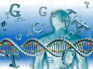
Nucleic Acids DNA and RNA is found in the nucleus of cells!!!! • There are TWO types of nucleic acids in living organisms: • DNA(DeoxyriboNucleic Acid) – the instructions for creating an organism are stored in digital code along coiled chains of DNA • RNA(RiboNucleic Acid) – reads the information in DNA and transports it to the protein-building apparatus of the cell
Nucleotide • Each nucleotide is made up of: • A 5 carbon sugar • a phosphate group • a nitrogenous base DNA is slightly acidic and composed of large amounts of phosphorus and nitrogen. The phosphate group is negatively charged.
Directionality of DNA nucleotide • You need to number the carbons! • it matters! PO4 N base 5 CH2 This will beIMPORTANT!! O 1 4 ribose 3 2 OH
DNA v. RNA • DNA contains the sugar DEOXYRIBOSE whereas RNA contains the sugar RIBOSE. The only difference between these two sugars is the lack of oxygen at carbon 2 in deoxyribose – this accounts for its name. DNA RNA
DNA nitrogen bases Purines have a double-ringed structure. Pyrimidines have a single-ringed structure.
RNA nitrogen bases • PURINES • 1. Adenine (A) • 2. Guanine (G) • PYRIMIDINES • 3. Uracil (U) • 4. Cytosine (C) Which base is found in DNA that is NOT found in RNA?
Erwin Chargaff 1947 • DNA composition: “Chargaff’s rules” • varies from species to species • all 4 bases not in equal quantity • bases present in characteristic ratio • humans: A = 30.9% T = 29.4% G = 19.9% C = 19.8% RulesA = T C = G That’s interesting!What do you notice?
DNA Structure “Legs of ladder” “Rungs of ladder” Nitrogen Base (A,T,G or C) Phosphate & Sugar Backbone Double Helix 1 main function ENERGY STORAGE short-term
Covalent bond: phosphodiester bond Hydrogen bonds 5 O 3 T A C G 3 O P P 5 5 O 3 1 2 4 4 1 2 3 5 O P P 3 5 O O 5 P P 3
Chargaff’s Rule C T A G • Adeninemust pair with Thymine • Guanine must pair with Cytosine
Nucleotide Pairing Complimentary base pairing causes the diameter of the DNA helix to remain constant ie: 2nm. The large number of hydrogen bonds between DNA strands account for its structural stability.
Antiparallel strands The phosphate group of one nucleotide is linked to the sugar of another via a PHOSPHODIESTER BOND. The end that terminates in a PHOSPHATE GROUP is labelled the 5’ end. The end that terminates in a HYDROXYL GROUP is labelled the 3’ end. Complimentary strands are said to be ANTIPARALLEL. The bond that links the sugar to its nitrogenous base is referred to as a GLYCOSYL BOND.
Nucleic Acids A P P P P P P C G G T A P 5’ P P P P P G C C A T T Hydrogen bonds 3’
Histones • Eukaryotic cells contain more than 2 m of DNA (over 6 billion base pairs) • DNA is wound twice around 8 histone proteins with an additional histone holding it all together. • This is called a nucleosome • This is further wound into chromatin fibres
DNA – double helix • Rosalind Franklin and Maurice Wilkins used X-ray diffraction to determine structure of DNA. Pattern was presented to James Watson and Francis Crick.
1953- Watson and Crick built model of DNA using work of Chargaff and Rosalind Franklin • Nobel prize- 1962 – Watson, Crick, Wilkins • Proposed right-handed model of DNA i.e. turns in clockwise direction • Makes one complete turn every 10 base pairs.
Sugar + phosphate = backbone • 2 strands run antiparallel i.e. one strand runs 5’ to 3’ – other strand 3’ to 5’ • i.e. 5’-ATGCCGTTA-3’ 3’-TACGGCAAT-5’ This is called complimentary base pairing
Structure of DNA 1953 | 1962 • Watson & Crick • developed double helix model of DNA • other leading scientists working on question: • Rosalind Franklin • Maurice Wilkins • Linus Pauling Wilkins Pauling Franklin
Scientific History T.H. Morgan (1908): genes are on chromosomes Frederick Griffith (1928): a transforming factor can change phenotype Avery, McCarty & MacLeod (1944): transforming factor is DNA Erwin Chargaff (1947): Chargaff rules: A = T, C = G Hershey & Chase (1952): confirmation that DNA is genetic material Watson & Crick (1953): determined double helix structure of DNA Meselson & Stahl (1958): semi-conservative replication