Download
1 / 92
1.06k likes | 1.45k Vues
Anatomy of the Cerebral Ventricles. Francois du Toit Diagnostic Radiology Kimberley Hospital. The Cerebral Ventricles. Fluid filled (CSF) spaces within the brain 2 Lateral ventricles in each hemisphere 3 rd ventricle, Cerebral A quaduct , 4th ventricle midline

E N D
Anatomy of the Cerebral Ventricles Francois du Toit Diagnostic Radiology Kimberley Hospital
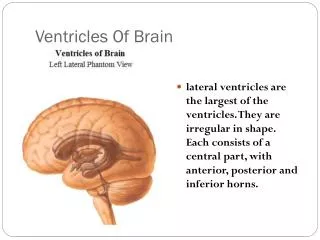
The Cerebral Ventricles • Fluid filled (CSF) spaces within the brain • 2 Lateral ventricles in each hemisphere • 3rd ventricle, Cerebral Aquaduct, 4th ventricle midline • Ependyma(thin epithelial membrane lining)
The Lateral Ventricles • Lies within each cerebral hemisphere: • Frontal Horn (anterior) • Body (atrium) • Temporal Horn (inferior) • Occipital Horn (posterior) • Interventricular Foramen (of Monroe) • connects each lateral ventricle with the 3rd ventricle • at junction of anterior horn & body
THE LATERAL VENTRICLE FRONTAL HORN
Frontal (anterior) Horn • Extends into frontal lobe • Roof & Ant extremity formed by: • Corpus Collosum (Rostrum & Genu) • Tapetum • Floor & Lateral Wall • Caudate Nucleus • Medial Wall • Septum Pellucidum
Frontal (anterior) Horn • Extends into frontal lobe • Roof & Ant extremity formed by: • Corpus Collosum (Rostrum & Genu) • Tapetum • Floor & Lateral Wall • Caudate Nucleus • Medial Wall • Septum Pellucidum
Frontal (anterior) Horn • Extends into frontal lobe • Roof & Ant extremity formed by: • Corpus Collosum (Rostrum & Genu) • Tapetum • Floor & Lateral Wall • Caudate Nucleus • Medial Wall • Septum Pellucidum
THE LATERAL VENTRICLE BODY (atrium)
Body of Lateral Ventricle • In Parietal Lobe • Roof & Lateral Wall • Corpus Callosum • TapetalFibres • Medial wall • Septum Pellucidum • Floor (medial) • Thalamus • Floor (lateral) • Body of Caudate Nucleus • Thalamostriatevein in between • Body of Fornix lies above the thalamus • Between the fornix and the thalamus • Choroid plexus lies invaginated into the cavity of the ventricle in a groove - the choroidal fissure
Body of Lateral Ventricle • In Parietal Lobe • Roof &Lat • Corpus Callosum • TapetalFibres • Medial wall • Septum Pellucidum • Floor (medial) • Thalamus • Floor (lateral) • Body of Caudate Nucleus • Thalamostriatevein in between • Body of Fornix lies above the thalamus • Between the fornix and the thalamus • Choroid plexus lies invaginated into the cavity of the ventricle in a groove - the choroidal fissure
Body of Lateral Ventricle • In Parietal Lobe • Roof &Lat • Corpus Callosum • TapetalFibres • Medial wall • Septum Pellucidum • Floor (medial) • Thalamus • Floor (lateral) • Body of Caudate Nucleus • Thalamostriatevein in between • Body of Fornix lies above the thalamus • Between the fornix and the thalamus • Choroid plexus lies invaginated into the cavity of the ventricle in a groove - the choroidal fissure
Body of Lateral Ventricle Caudate Nucleus Thalamus
Body of Lateral Ventricle • In Parietal Lobe • Roof &Lat • Corpus Callosum • TapetalFibres • Medial wall • Septum Pellucidum • Floor (medial) • Thalamus • Floor (lateral) • Body of Caudate Nucleus • Thalamostriatevein in between • Body of Fornix lies above the thalamus • Between the fornix and the thalamus • Choroid plexus lies invaginated into the cavity of the ventricle in a groove - the choroidal fissure
The Lateral Ventricles Body of Fornix Choroid Plexus Thalamus
THE LATERAL VENTRICLE TEMPORAL HORN (inferior)
Temporal (inferior) Horn • Extends anteriorly into Temporal Lobe • Lateral wall • Fibresof Tapetum • Roof • Tail of Caudate Nucleus • Floor • Hippocampus • peshippocampi anterior • crus of the fornix arising from this
Tapetum Temporal Horn of Lateral Ventricle
Temporal (inferior) Horn • Extends anteriorly into Temporal Lobe • Lateral wall • Fibresof Tapetum • Roof • Tail of Caudate Nucleus • Floor • Hippocampus • peshippocampi anterior • crus of the fornix arising from this
Temporal (inferior) Horn • Extends anteriorly into Temporal Lobe • Lateral wall • Fibresof Tapetum • Roof • Tail of Caudate Nucleus • Floor • Hippocampus • peshippocampi anterior • crus of the fornix arising from this
Crus of Fornix Hippocampus Pes
THE LATERAL VENTRICLE OCCIPITAL HORN (Posterior)
Occipital (posterior) Horn • Posterior Extension of Lateral ventricle • Extends into Occipital Lobe • Arises from trigone of lateral ventricle • posterior convexity of the body of the lateral ventricle • May be absent / poorly developed / extend the full depth • 12% bilaterally well developed
Choroid Plexus of Lateral Ventricle • Responsible for most of the production of CSF • Extends from Inferior horn, through body, to interventricularforamen • NO CHOROID PLEXUS in Occipital & Frontal Horn • Continuous with Choroid Plexus of 3rd ventricle • Invaginatedinto Lateral Ventricles medially (Choroidal Fissure)
Choroid Plexus of Lateral Ventricle • Blood supply: • Ant Choroidal a. (Branch of ICA) • Post Choroidal a. (Branch of post Cerebral a.) • Venous drainage: • Sup Choroidalvein (begins at inferior horn and passes anteriorly to IV foramen) • Joins Sup Thalamostriatev. to form Internal Cerebral vein at IV foramen
3rdVentricle • Slit-like space between Thalami • Width = 2-10mm (increasing with age) • Thin anterior wall – Lamina Terminalisbetween ant commissure (above) to optic chiasm (below) • Extension inferiorly into optic chiasm = supraoptic recess • Floor = Structures of hypothalamus including pituitary whose hollow stalk is the infundibular recess of the ventricle • Fold of Pia containing CP = TelaChoroidea • Narrow Anterior Apex at IV Foramen • Wider Posterior • If Fluid accumulates = Cavum Velum Interpositum
Thalamus Third Ventricle
3rdVentricle • Slit-like space between Thalami • Width = 2-10mm (increasing with age) • Thin anterior wall – Lamina Terminalisbetween ant commissure (above) to optic chiasm (below) • Extension inferiorly into optic chiasm = supraoptic recess • Floor = Structures of hypothalamus including pituitary whose hollow stalk is the infundibular recess of the ventricle • Fold of Pia containing CP = TelaChoroidea • Narrow Anterior Apex at IV Foramen • Wider Posterior • If Fluid accumulates = Cavum Velum Interpositum
Anterior Commisure Lamina Terminalis Optic Chiasm
3rdVentricle • Slit-like space between Thalami • Width = 2-10mm (increasing with age) • Thin anterior wall – Lamina Terminalisbetween ant commissure (above) to optic chiasm (below) • Extension inferiorly into optic chiasm = supraoptic recess • Floor = Structures of hypothalamus including pituitary whose hollow stalk is the infundibular recess of the ventricle • Fold of Pia containing CP = TelaChoroidea • Narrow Anterior Apex at IV Foramen • Wider Posterior • If Fluid accumulates = Cavum Velum Interpositum
3rdVentricle • Slit-like space between Thalami • Width = 2-10mm (increasing with age) • Thin anterior wall – Lamina Terminalisbetween ant commissure (above) to optic chiasm (below) • Extension inferiorly into optic chiasm = supraoptic recess • Floor = Structures of hypothalamus including pituitary whose hollow stalk is the infundibular recess of the ventricle • Fold of Pia containing CP = TelaChoroidea • Narrow Anterior Apex at IV Foramen • Wider Posterior • If Fluid accumulates = Cavum Velum Interpositum
3rdVentricle • Slit-like space between Thalami • Width = 2-10mm (increasing with age) • Thin anterior wall – Lamina Terminalisbetween ant commissure (above) to optic chiasm (below) • Extension inferiorly into optic chiasm = supraoptic recess • Floor = Structures of hypothalamus including pituitary whose hollow stalk is the infundibular recess of the ventricle • Fold of Pia containing CP = TelaChoroidea • Narrow Anterior Apex at IV Foramen • Wider Posterior • If Fluid accumulates = Cavum Velum Interpositum