Download
1 / 26
320 likes | 542 Vues
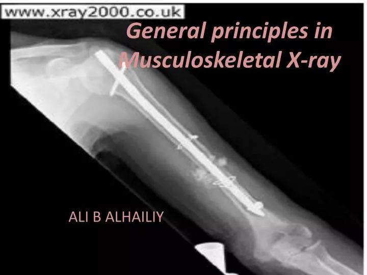
General principles in Musculoskeletal X-ray. ALI B ALHAILIY. Definition. A fracture is present when there is loss of continuity in the substance of a bone. The term covers all bony disruptions, ranging from the situation when a bone is broken into one or many fragments.

E N D
General principles in Musculoskeletal X-ray ALI B ALHAILIY
Definition • A fracture is present when there is loss of continuity in the substance of a bone. • The term covers all bony disruptions, ranging from the situation when a bone is broken into one or many fragments
Causes of fracture • Fractures are caused by the application of stresses which exceed the limits of strength of a bone. • Violence is the commonest cause • 1- direct violence • abone may be fractured by being struck by a moving or falling object. • 2- indirect violence. • twisting or bending stress is applied to a bone, and this results in its fracture at some distance from the application of the causal force.
Types of fracture • 1-Pathological fracture • Sometimes fracture may occur in an abnormal or diseased bone. • That will reduce the strength of the bone then the force required to produce fracture is reduced. Bone cyst
2-Fatigue fractures (stress fractures) • Stresses, repeated with excessive frequency to a bone, may result in fracture. • It is hairline in pattern and are often not diagnosed with certainty until there iscallus formation, or increased density at the fracture site. • 3-6 weeks after the onset of symptoms Healed fatigue fracture through the second metatarsal bone
3- Open and closed fractures • Closed fracture: (a) the bone is • broken, but there is no external wound. • A fracture not communicating with the external environment • The overlying skin are intact • Open fracture: A fracture with break in the overlying skin and soft-tissues, • leading to the fracture.
4 4- Simple transveres fractures • Transverse fractures run either at right angles to the long axis of a bone (1), or with an obliquity of less than 30°. • 5-Multifragmentalfractures • There are two fragments or more. 5
6- Avulsion fractures : produced by a sudden muscle contraction • the muscle pulling off the portion of bone to which it is attached. • Common examples include: • (1) Base of fifth metatarsal. • (2) Tibialtuberosity (quadriceps). • (3) Upper pole of patella (quadriceps). • (4) Lesser trochanter (iliopsoas).
7-Fracture-dislocations • present when a joint has dislocated and there is in addition a fracture of one of the bony components of the joint. • fracture-dislocation of the shoulder, where there is an anterior dislocation with a fracture of the neck of the humerus. • Injuries of this kind may be difficult to reduce and may be: • unstable. Stiffness and avascular necrosis are two common complications
Describing the level of a fracture • The anatomical divisions of a long bone include: • the epiphysis (E), epiphyseal plate (EP), and diaphysis or shaft (D). Between the latter two is the metaphysis (M). • A fracture may be described as lying within these divisions, or involving a distinct anatomical part, e.g. • A = fracture of the tibialdiaphysis OR ????? • B = fracture of the femoral neck; • C = fracture of the greater trochanter; • F = supracondylar fracture of the femur OR ?????????????????????.
Describing the level of a fracture • For descriptive purposes a bone may be divided into thirds. In this way: • A = fracture of the mid third of the femur; • B = fracture of the femur in the distal third; • C = fracture of the femur at the junction of the • middle and distal thirds. • D = fracture of the distal metaphysis of radial bone
Musculoskeletal X-ray - General principles • Please have on the systemic approach and viewing principles tutorials at: • http://radiologymasterclass.co.uk/tutorials/musculoskeletal/principles/bones_joints_x-ray_page4.html
X RAY FINDINGS Phalangeal dislocations Forced hyperextension of the thumb or finger joints is the usual cause for these injuries. Normally the distal segment is displaced posteriorly
Bennett’s fracture • It is a fractureof the base of the first metacarpal bone which extends into the carpometacarpal(CMC) joint. • This intra-articularfracture is the most common type of fracture of the thumb • A vertical lucent line is seen intersecting the proximal articular surface of the first metacarpal bone
Scaphoid Fracture • Usually fractures through the waist (middle) of the scaphoid bone • Mechanism – fall on dorsiflexed outstretched hand • May not be evident for up to 10 days post trauma • Very well diagnosed clinically – pain and tenderness in anatomical snuff box • Poor healing fracture due to complicated blood supply • Most easily diagnosed via the PA ulnar deviation with angulation projection
Scaphoid fractures • How many projections for scaphoid # ?
Or… PA OBL PA PA DEV LAT General setup of 24 x 30 cm cassette for patients who have defined trauma to scaphoid or follow up films or minimal injury
Colles’ Fracture • # of the distal end of the radius (3cm from joint) +/- # of the styloid process of the ulna • Dorsal angulation of the distal fragment – ventral angulation of proximal radius • Most common form of wrist fracture – females, middle aged and older • Caused by fall on outstretched hand (FOOSH)
Smith’s Fracture • Reverse Colles’ # • Hyperflexion with fall on back of hand • Fracture of the radius with ventral displacement of distal fragment and dorsal angulation of proximal radius
Galeazzi’s Fracture • # of the mid shaft of the radius • Dislocation of the distal radioulnar joint • Fall on outstretched hand with elbow flexed • High incidence of nonunion and a limitation on pronating or supinating the hand
Please read about • Salter–Harris fractures ? • Types with figures ? • http://cal.vet.upenn.edu/projects/saortho/chapter_11/11mast.htm