Download
1 / 38
390 likes | 450 Vues
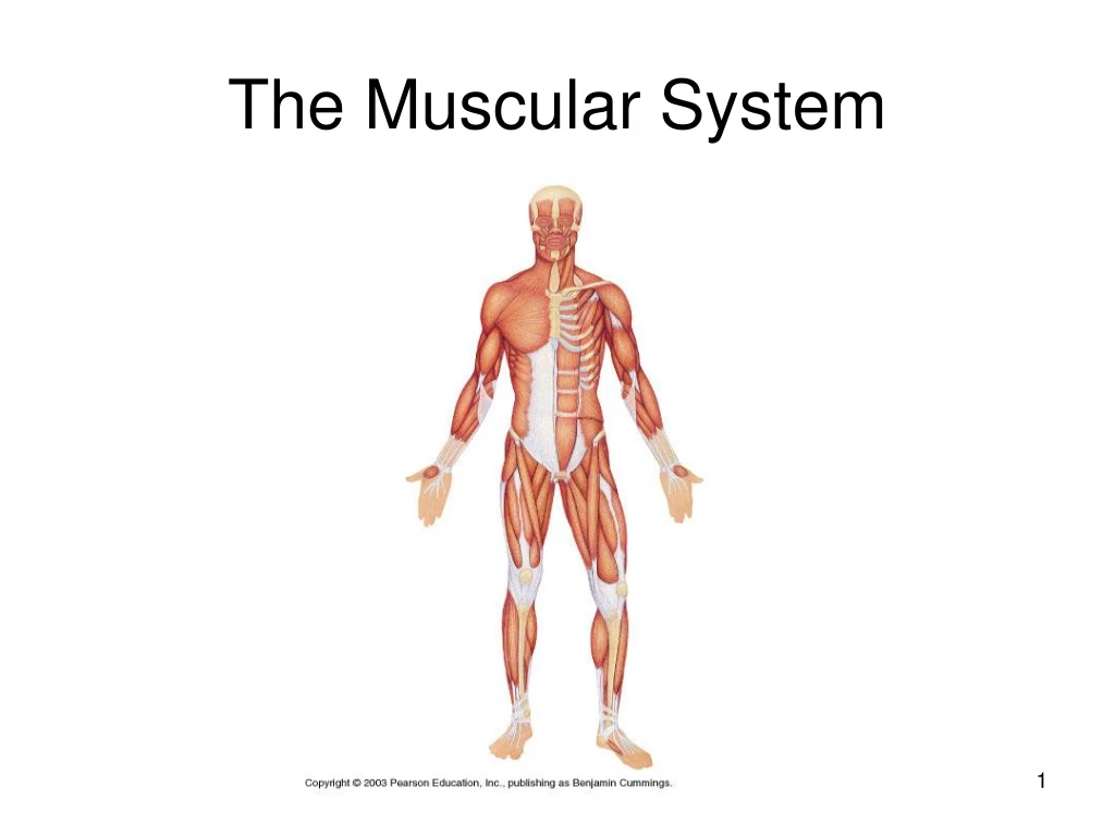
The Muscular System. Objectives:. List and describe the major functions of the muscular system Describe the structure of a skeletal muscle at the macroscopic and microscopic level Describe muscle contraction according to the sliding-filament theory

E N D
Objectives: • List and describe the major functions of the muscular system • Describe the structure of a skeletal muscle at the macroscopic and microscopic level • Describe muscle contraction according to the sliding-filament theory • Name and identify the location of major muscles and muscle groups of the body • List and describe diseases and disorders of the muscular system.
Function of Muscles • Produce movement • Maintain posture • Stabilize joints • Generate heat
Characteristics of Muscles • Muscle cells are elongated (muscle cell = muscle fiber) • Contraction of muscles is due to the movement of microfilaments • All muscles share some terminology • Prefix myo refers to muscle • Prefix mys refers to muscle • Prefix sarco refers to flesh • Most are attached by tendons to bones • Striated – have visible banding • Cells are surrounded and bundled by connective tissue
Macroscopic Structure • Fascia – on the outside of the epimysium, it is the hypodermis • Epimysium – covers the entire skeletal muscle • Perimysium – around a fascicle (bundle) of fibers • Endomysium – around single muscle fiber • Origin - the point at which the muscle attaches to a structure to provided resistance to create movement. • Insertion – the point at which the muscle attaches to the structure which is moved when it contracts.
Microscopic Structure of Myofibril • Cells are multinucleate • Nuclei are just beneath the sarcolemma • Sarcolemma – specialized plasma membrane • Sarcoplasmic reticulum – specialized smooth endoplasmic reticulum which stores calcium ions
Microscopic Structure of Myofibril • Myofibril • Bundles of myofilaments • Myofibrils are aligned to give distinct bands • I band = light band • A band = dark band • Sarcomere • Contractile unit of a muscle fiber, which extends from one z line or disc to the next z line or disc.
Microscopic Structure of Sarcomere • Organization of the sarcomere • Thick filaments = myosin filaments • Composed of the protein myosin • Has ATPase enzymes • Thin filaments = actin filaments • Composed of the protein actin • Has troponin and tropomyosin components I Band A Band I Band
Microscopic Structure of Myofilaments • Myosin filaments have heads (extensions, or cross bridges) • Myosin and actin overlap somewhat • At rest, there is a bare zone that lacks actin filaments this is the location of the M line and the H zone.
Properties of Muscle Fibers Which Produce Movement • Irritability – ability to receive and respond to a stimulus • Contractility – ability to shorten when an adequate stimulus is received • Extensibility- ability to lengthen when it is relaxed and not being stimulated
How Muscle Fibers Produce Movement • to produce movement, skeletal muscles must be stimulated by a motor neuron • Motor unit • One neuron • Neuromuscular junctions – association site of nerve and muscle
Synaptic cleft – gap between nerve and muscle Nerve and muscle do not make contact Area between nerve and muscle is filled with interstitial fluid Neurotransmitter – chemical released by nerve upon arrival of nerve impulse The neurotransmitter for skeletal muscle is acetylcholine Ach attaches to receptors on the sarcolemma Sarcolemma becomes permeable to sodium (Na+) Neuromuscular Junction
Neuromuscular Junction • Sodium rushing into the cell generates an action potential. • Once this happens, the sarcoplasmic reticulum releases calcium ions. • This begins the contraction of the sarcomere units in the myofibrils. • Once started, the contraction of the muscle fiber cannot be stopped (All or none response- either the entire muscle fiber contracts or it does not contract at all.)
Sliding Filament Theory Relaxed Sarcomere • ATP on the myosin filament causes the myosin head to move from the relaxed state to the upright excited state • release of calcium ions expose the binding sites on actin filaments to which the heads of the myosin filaments bind • Myosin heads then bind to the exposed site of the Actin (troponin/tropomyosin) • Once the crossbridges form, the myosin head bends towards the M line or H zone. • This continued action causes a sliding of the myosin along the actin pulling the actin filament and z lines toward the center or H zone. The H zone disappears. • The result is that the muscle is shortened (contracted) Contracted Sarcomere
Sliding Filament Theory Tropomyosin Troponin
Muscle Contraction • Muscle force depends upon the number of fibers stimulated • Within a skeletal muscle, not all fibers may be stimulated during the same interval • Different combinations of muscle fiber contractions may give differing responses • More fibers contracting results in greater muscle tension • Muscles can continue to contract unless they run out of energy
Pop Quiz 1. 4. Is this muscle relaxed or contracted? 2. Key Terms: Myofibril Sarcomere Myosin Actin 5. Is this muscle relaxed or contracted? How do you know? 3. The area represented by letter A is called the ____.
Types of Muscle • Prime mover – muscle with the major responsibility for a certain movement • Antagonist – muscle that opposes or reverses a prime mover • Synergist – muscle that aids a prime mover in a movement and helps prevent rotation • Fixator – stabilizes the origin of a prime mover
Effects of Exercise on Muscles • Results of increased muscle use • Increase in muscle size (The mass of the muscle fibers increases, not an increase in muscle fiber number!) • Increase in muscle strength • Increase in muscle efficiency • Muscle becomes more fatigue resistant
Naming of Muscles • Direction of muscle fibers • Example: rectus (straight) • Relative size of the muscle • Example: maximus (largest), major (larger of group) • Location of the muscle • Example: many muscles are named for bones (e.g., temporalis) • Number of origins • Example: triceps (three heads) • Location of the muscles origin and insertion • Example: sterno (on the sternum) • Shape of the muscle • Example: deltoid (triangular) • Action of the muscle • Example: flexor and extensor (flexes or extends a bone)
Pop Quiz 1 Key Choices Occipitalis Masseter Temporalis Orbicularis occuli Platysma Frontalis Orbicularis oris Zygomaticus 4 2 5 6 3
Diseases and Disorders of the Muscular System • Cerebral Palsy: This disorder is characterized by paralysis and or weakened muscles due to loss of muscle tone. It can be caused due to lack of oxygen to the motor region of the cerebrum of the brain which controls conscious control of muscles. This is often attributed to complication during birth.
Diseases and Disorders of the Muscular System • Myalgia: Muscle pain due to strain, tearing of muscle fibers. It also is a symptom of an immune response along with a fever. • Myositis: Inflammation of muscle tissue due to injury or disease. • Charley Horse (fibromyositis): Inflamation of muscle tissue and the tendons associated with that muscle due to injury (tear or severe bruising- contusion) • Cramps: Painful, involuntary muscle spasms
Diseases and Disorders of the Muscular System • Poliomyelitis: Polio is due to a viral infection which affects the motor neurons that control skeletal muscles. It often leads to paralysis and can result in death by paralysis of the diaphragm. Due to vaccine developed by Jonas Salk, the virus has been virtually eliminated in the US. However, it still poses a threat in developing countries.
Diseases and Disorders of the Muscular System • Muscular Dystrophy: Series of genetic disorders characterized by the atrophy or wasting away of skeletal muscle. Duchenne Muscular Dystrophy is the most common and affects primarily males. The muscle tissue breaks down and is replaced by fat and fibrous tissue. DMD is characterized by weakness in the leg muscles which then rapidly spreads to the shoulders and other parts of the body. Death usually occurs by the age of 21 due to respiratory or cardiac muscleweakness.
Functions Microscopic and macroscopic anatomy of muscles Muscle physiology – neuromuscular junction and sliding filament theory (muscle contraction process) Types of body movements 4 Types of muscles Naming of muscles Major muscles diagrams Facial muscles diagrams and definitions Diseases of muscular system P. 198 – 199 MC #1,2,4,5 SA #1-7, 10-13 Study Outline