Download
1 / 64
640 likes | 812 Vues
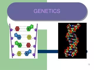
GENETICS. CONTENTS. 1. Diagnostic techniques, M utations , Chromosome disorders 2 . Mendelian d isorders , Multifactorial inheritance, M itochondrial DNA D isorders 3 . Sex differentiation disorders, Developmental disorders. Diagnostic techniques, Mutations, Chromosome disorders.

E N D
CONTENTS • 1. Diagnostic techniques, Mutations, Chromosome disorders • 2. Mendeliandisorders, Multifactorialinheritance, Mitochondrial DNA Disorders • 3. Sex differentiation disorders, Developmental disorders
What are the most common genetic disorders in blacks • Sickle cell trait/disease, a- and b-thalassemia, Glucose-6-phosphate dehydrogenase (G6PD) deficiency • What are the most common genetic disorders in Jews • Tay-Sachs disease, Gaucher's disease, factor XII deficiency • What are the most common genetic disorder in Northern Europeans • Cystic fibrosis
What are the most common types of prenatal tests • Amniocentesis (16 weeks), chorionic villus biopsy (9-12 weeks), fetal blood sampling, fetal biopsy, ultrasonography • What are the most common routine maternal screening tests for genetic disorder • Alpha-fetoprotein, beta-human chorionic gonadotrophin, unconjugatedestriol [A low alpha-fetoprotein and unconjugatedestriol + a high (beta-human chorionic gonadotropin occurs in Down syndrome in 60% of cases.] • What are the most common screening test abnormality in open neural tube defects • Increased alpha-fetoprotein [alpha-fetoprotein is fetal albumin. It must be correlated with gestational age.]
What are the most common DNA technique used to locate gene abnormalities • Nucleic acid probes [Probes contain specific amino acid sequences of portions of DNA. They are spliced into DNA strands and are allowed to hybridize with a corresponding segment of DNA from a patient sample.] • What are the most common cause of Down syndrome • Nondisjunction resulting in trisomy 21 (extra chromosome is of maternal origin) [Increasing maternal age increases the risk for non-disjunction.] • What are the most common cause of death in childhood in Down syndrome • Endocardial cushion defects (combined Atrialseptal defect and Ventricular septal defect)
What are the most common factor affecting longevity in older patients with Down syndrome • Alzheimer's disease [Chromosome 21 codes for (3-amyloid protein, which is converted into amyloid, which is toxic to neurons.] • What are the most common Gastrointestinal abnormalities in Down syndrome • Duodenal atresia (vomiting of bile-stained material at birth; double bubble sign on a flat plate of the abdomen) and Hirschsprung's disease (absent ganglion cells in the rectum; failure to pass meconium) • What are the most common hematologic problem in Down syndrome • Acute myelogenous leukemia (<3 years of age) and acute lymphoblastic leukemia (>3 years of age)
What are the most common risk factor for having a child with Down syndrome • Maternal age [Any woman >35 years of age is at increased risk.] • What are the most common genetic cause of primary amenorrhea [no menses by 16 years of age] • Turner's syndrome [Owing to the absence of oocytes by 2 years of age, these patients have decreased estradiol levels, hence they lack secondary sex characteristics and do not menstruate.] • What are the most common cause of Turner's syndrome • Nondisjunction leading to a 45 XO genotype (-50-60%) [Mosaics may also occur (XO/XY; XO/XX). Since one of the two X chromosomes is randomly inactivated to become a nuclear appendage called a Barr body, Turner's syndrome patients do not have any Barr bodies (a normal woman has one Barr body).]
What are the most common study used to identify Barr bodies • Smear of the buccal mucosa squamous cells and identification of nuclear appendages representing Barr bodies • What are the most common newborn presentation of Turner's syndrome • Lymphedema of the hands and feet, cystic hygroma (dilated lymphatic channels) in the neck, preductalcoarctation of the aorta • What are the most common ovarian tumor associated with Turner's syndrome • Dysgerminoma (germ cell tumor) [Presence of a Y chromosome confers an increased risk for ovarian cancer.]
What are the most common lab findings in Turner's syndrome • Decreased estradiol, increased Follicle-stimulating hormone and Luteinizing-hormone levels [Decreased estradiol results in an increase in gonadotropins owing to a negative feedback relationship.] • What are the most common cause of Klinefelter's syndrome • Nondisjunction resulting in an XXY genotype [In contrast to a normal male, there is one Barr body.] • What are the most common testicular findings in Klinefelter's syndrome • Atrophy with fibrosis of the seminiferous tubules and hyperplasia of Leydig cells
What are the most common cause of hyperestrinism in Klinefelter's syndrome • Leydigcells [SerAromatization of testosterone into estradiol in the tolicells in the seminiferous tubules normally synthesize inhibin, an inhibitor of Follicle-stimulating hormone. Absence of inhibin increases Follicle-stimulating hormone, which increases the synthesis of aromatase in Leydig cells. Patients have female secondary sex characteristics (e.g., gynecomastia).] • What are the most common lab findings in Klinefelter's syndrome • Decreased testosterone, azoospermia (no sperm), increased Follicle-stimulating hormone, Leutinizing-hormone, and estradiol • What are the most common signs and symptoms of the cri-du-chat syndrome • Mental retardation, cat-like cry, and a Ventricular septal defect (due to a partial deletion of chromosome 5)
What are the most common microdeletion syndromes • Prader-Willi and Angelman's syndromes [Both have a microdeletion at the same location on chromosome 15, but in the former syndrome, chromosome 15 is of paternal origin, and in the latter syndrome, it is of maternal origin. This is an example of genomic imprinting.]
Mendelian disorders, Multifactorial inheritance, Mitochondrial DNA Disorders
What are the most common Mendelian disorder • Autosomal dominant (AD) [Dominant means that the gene is strong enough to express itself in a heterozygous state (one normal allele and one abnormal allele; an allele is an alternative form of the same gene). Homozygous Autosomal dominant diseases are rare. 50% of siblings are affected and 50% are normal when one of the parents is normal and the other has an Autosomal dominant disease.]
What are the most common characteristics of Autosomal recessive diseases • Recessive indicates a weak gene that must be present in a homozygous state (both alleles are abnormal) for it to express itself; heterozygotes are asymptomatic carriers [Both parents must be carriers of the abnormal gene to transmit the disease to their children. Two asymptomatic carriers have a 25% chance of having a child with the disease, a 50% chance of a child who is an asymptomatic carrier, and a 25% chance of a normal child,
What are the most common characteristics of Sex-linked Recessive disorders • An affected male transmits the disease to all his daughters, who are asymptomatic carriers, and to none of his sons. An asymptomatic carrier female transmits the disease to 50% of her sons and 50% of her daughters, the latter representing asymptomatic carriers
What are the most common characteristics of Sex-linked Dominant disorders • The percentages of children with the abnormal gene are the same as with Sex-linked recessive; however, the gene is dominant, so both males and females are symptomatic [An affected woman transmits symptomatic disease to 50% of her daughters and 50% of her sons and an affected man transmits disease to all of his daughters and none of his sons.] • What are the most common Mendelian disorder exhibiting penetrance • Autosomal Dominant [Penetrance is where a person with the abnormal gene may or may not express the disease. If two children are Aa but only one of the two expresses the disease, there is 50% penetrance (100% penetrance if both express the disease). The asymptomatic person can still transmit the disease to his or her children.]
What are the most common Mendelian disorder exhibiting variable expressivity • Autosomal Dominant [Variable expressivity is where there can be varying degrees of expression of the disease. Unlike with penetrance, the patient does manifest the disease to some minor or major extent.] • What are the most common Mendelian disorder in which the disease may occur later in life • Autosomal Dominant [Examples of this phenomenon are Huntington's chorea, adult polycystic kidney disease, and familial polyposis. Chorea, renal cysts, and polyps, respectively, are not present at birth.] • What are the most common Autosomal Dominant disease • von Willebrand's disease (1 in 125)
What are the most common Autosomal Recessive disease • Hemochromatosis (1 in 100) • What are the most common Sex-linked Recessive disease • Fragile X syndrome (1 in 1000) • What are the most common genetic disease in blacks • Sickle cell disease (1 in 625)
What are the most common Mendelian disorder associated with enzyme deficiencies • Autosomal Recessive [A few Sex-linked Recessive diseases have enzyme deficiencies (e.g., Glucose-6-Phosphate dehydrogenase deficiency, Lesch-Nyhan syndrome), and rarely, Autosomal Dominant diseases (e.g., Acute intermittent porphyria, Cl esterase inhibitor deficiency).] • What are the most common Mendelian disorder associated with structural defects in proteins and receptors • Autosomal Dominant [For example, congenital spherocytosis has a defect in spectrin in the RBC membrane. Familial hypercholesterolemia has a defect in the Low-density lipoprotein receptor.]
What are the most common skin lesions associated with neuroflbromatosis • Autosomal Dominant disease with cafe au lait spots; pigmented, pedunculatedneurofibromas; and plexiformneurofibromas [Cafe au lait spots are coffee-colored flat lesions whose long axes parallel an underlying cutaneous nerve. Pedunculatedneurofibromas are benign tumors that involve all elements of the peripheral nerve. Plexiformneurofibromas lead to grotesque overgrowth of soft tissue (e.g., "elephant man").] • What are the most common tumors associated with neurofibromatosis • Acoustic neuromas (benign tumor arising from Schwann cells involving the eighth cranial nerve), meningiomas, optic nerve gliomas, pheochromocytomas (produces hypertension), and Lisch nodules on the iris (really hamartomas, not true neoplasms)
What are the most common defect in Marfan's syndrome (Autosomal Dominant) • Defect in fibrillin, a component of elastin in elastic tissue • What are the most common skeletal defects in Marfan's syndrome • Eunuchoid proportions (lower body length greater than upper body length; arm span greater than height), arachnodactyfy (spider hands) • What are the most common cardiovascular abnormalities in Marfan's syndrome • Dissecting aortic aneurysm (most common cause of death), mitral valve prolapse (common cause of sudden death)
What are the most common eye defect in Marfan's syndrome • Dislocated lens [The suspensory ligament is defective.] • What are the most common enzyme deficiency, in Phenylketonuria (Autosomal Recessive) • Phenylalanine hydroxylase deficiency, which catalyzes the conversion of phenylalanine into tyrosine [Phenylalanine is increased and tyrosine is decreased. A blood sample must be taken a few days after birth and not on cord blood, since the baby must be exposed to phenylalanine in breast milk.] • What are the most common signs and symptoms in Phenylketonuria • Mental retardation, projectile vomiting (resembles congenital pyloric stenosis), hypopigmentation (decreased tyrosine = decrease in melanin synthesis)
What are the most common buildup products in Phenylketonuria • Phenylalanine, phenylpyruvic acid, phenyl-lactic acid, and phenylacetic acid [The last three compounds are minor pathway products derived from phenylalanine. They impart a mousy odor to sweat.] • What are the most common treatment for Phenylketonuria • Phenylalanine-free diet • What are the most common enzyme deficiency in galactosemia (Autosomal Recessive) • Galactose-1-phosphate uridyltransferase, which converts galactose 1-PO4 into glucose 1-PO4, the latter converted into glucose 6-PO4 (fuel for gluconeogenesis)
What are the most common signs and symptoms of galactosemia • Toxic effects on multiple organs and osmotic damage to the lens and peripheral nerves [An increase in galactose 1-PO4 is toxic to neurons (mental retardation), hepatocytes (liver failure), and the kidneys (aminoaciduria). Galactose is converted into galactitol, an alcohol sugar, which produces osmotic damage to the lens (cataracts) and peripheral nerve (peripheral neuropathy).] • What are the most common lab findings in galactosemia • Galactosuria, hypoglycemia [Galactose spills into the urine, producing a positive test result for reducing substances. The decrease in glucose 6-PO4 causes fasting hypoglycemia.]
What are the most common treatment for galactosemia • Lactose-free diet [Lactose is glucose + galactose.] • What are the most common enzyme deficiency in essential fructosuria (Autosomal Recessive) • Fructokinase, which catalyzes the reaction that converts fructose into fructose 1-PO4 [Unlike hereditary fructose intolerance, it is a benign disease with an excess of fructose in the urine.] • What are the most common enzyme deficiency in hereditary fructose intolerance (Autosomal Recessive) • Aldolase B [Aldolase B catalyzes the reaction that converts fructose 1-PO4 into glyceraldehyde and DHAP (dihydroxyacetone phosphate), which are three carbon intermediates in the glycolytic cycle.]
What are the most common signs and symptoms of hereditary fructose intolerance • A buildup of fructose 1-PO4, which is toxic to neurons (mental retardation) and hepatocytes (liver failure) • What are the most common lab findings in hereditary fructose intolerance • Fructosuria, hypophosphatemia, hypoglyce-mia [Fructose spills into the urine, producing a positive test result for reducing substances. Phosphorylation of fructose intracellularly depletes PO4 stores (hypophosphatemia). The decrease in three carbon intermediates reduces substrates for gluconeogenesis (fasting hypoglycemia) . ]
What are the most common enzyme deficiency in homocystinuria (Autosomal Recessive) • Cystathionesynthetase, which converts homocysteine into cystathione • What are the most common signs and symptoms of homocystinuria • Homocysteine is increased, leading to vessel damage and thrombosis [Other features resemble Marfan's syndrome (dislocated lens, arachnodactyly). Two diseases that are dissimilar to each other in pathogenesis but similar in clinical findings are an example of genetic heterogeneity.] • What are the most common enzyme deficiency in alkaptonuria (Autosomal Recessive) • Homogentisateoxidase, which converts homogentisate into maleylacetoacetate [Homogentisic acid is increased.]
What are the most common signs and symptoms of alkaptonuria • Homogentisic acid (oxidized into a black pigment) binds to collagen (severe joint disease, intervertebral disc disease) and produces black urine on exposure to light • What are the most common enzyme deficiency in von Gierke's disease (Autosomal Recessive) • It is a glycogen storage disease caused by a deficiency of the gluconeogenic enzyme glucose 6-phosphatase, which cacatalyzes the conversion of glucose 6-PO4 into glucose • What are the most common signs of von Gierke's disease • Hepatorenomegaly due to glycogen excess with liver and renal failure (gluconeogenic enzymes are in these organs) [Glucose 6-PO4 is the substrate for producing glycogen (glucose 6-PO4 -» glucose 1-PO4 -» UDP-glucose — » glycogen), hence its increase leads to an excess production of normal glycogen in organs containing the missing gluconeogenic hormones.]
What are the most common lab findings in von Gierke's disease • Hypoglycemia, lactic acidosis, hyperuricemia [Glucose cannot be synthesized in the fasting state (hypoglycemia) and there is an increase in anaerobic metabolism (lactic acidosis). Glucagon, cortisol, or fructose challenges do not increase glucose because of the enzyme block. Glycogen accumulation in the kidneys interferes with uric acid excretion.] • What are the most common enzyme deficiency in Pompe's disease (Autosomal Recessive) • It is the only glycogenosis that is a lysosomal storage disease, since it is deficient in the lysosomal enzyme acid maltase, which is necessary to degrade glycogen [Normal glycogen accumulates in the heart, leading to a restrictive cardiomyopathy at an early age.]
What are the most common enzyme deficiency in McArdle's disease (Autosomal Recessive) • It is a glycogenosis due to a deficiency of muscle phosphorylase, hence muscle glycogen cannot be catabolized into glucose for energy • What are the most common signs and symptoms of McArdle's disease • Patients fatigue easily and rhabdomyolysis (muscle necrosis) may occur [Lactic acid does not accumulate after exercise, since anaerobic glycolysis cannot occur without glucose.] • What are the most common types of glycogenoses associated with an accumulation of abnormal glycogen • Brancher and debrancher enzyme deficiency types of glycogenoses. [Von Gierke's disease, Pompe's disease, and McArdle's disease all have an accumulation of normal glycogen.]
What are the most common enzyme deficiency in Tay-Sachs disease (AR) • It is a sphingolipidosis (lysosomal storage disease) due to a deficiency of hexosaminidase leading to an accumulation of GM2 gangliosides in lysosomes (forms whorled configurations on Electron microscopy) [It is associated with a cherry red spot in the macula and severe mental retardation.] • What are the most common enzyme deficiency in Niemann-Pick disease (Autosomal Recessive) • It is a sphingolipidosis (lysosomal storage disease) with a deficiency of sphingomyelinase leading to an accumulation of sphingomyelin (forms zebra bodies on Electron microscopy) [Macrophages have a bubbly cytoplasm. Patients are mentally retarded and have hepatosplenomegaly.j
What are the most common enzyme deficiency in Gaucher's disease (Autosomal Recessive) • It is a sphingolipidosis (lysosomal storage disease) due to a deficiency of beta-glucocerebrosidase leading to an accumulation of glucocerebroside [Macrophages have a fibrillar-appearing cytoplasm. Hepatosplenomegaly is prominent.] • What are the most common enzyme deficiency in metachromaticleukodystrophy (Autosomal Recessive) • It is a sphingolipidosis (lysosomal storage disease) due to a deficiency of arylsulfatase A with an accumulation of sulfatides [An abnormal myelin is produced.] • What are the most common enzyme deficiency in Krabbe's disease (Autosomal Recessive) • It is a sphingolipidosis (lysosomal storage disease) due to a deficiency of galactosylceramidase and an accumulation of galactocerebroside [In the Central nervous system, the galactocerebroside is phagocytosed by histiocytes producing multinucleated giant cells called globoid bodies.]
What are the most common lysosomal storage diseases that are Sex-linked recessive • Fabry's disease and Hunter's syndrome [The former is due to a deficiency of a-galactocere-brosidase A with an accumulation of ceramidetrihexoside and the latter a deficiency of L-iduronosulfatesulfatase and an accumulation of dermatan and heparan sulfate.] • What are the most common mucopolysaccharidosis • Hurler's syndrome (Autosomal Recessive), which is due to a deficiency of a-1-iduronidase and an accumulation of dermatan sulfate and heparan sulfate [Patients have severe mental retardation, coarse facial features, premature Coronay artery disease, and abnormal circulating lymphocytes with vacuoles.]
What are the most common enzyme deficiency in Lesch-Nyhan syndrome (Sex-linked Recessive) • It is due to a deficiency of Hypoxanthine-guanine-phosphoribosyl-transferase, an inhibitor of 5-phospho-u-D-ribosyl-l-pyrophosphate (PRPP), hence purine metabolism is left unchecked and uric acid is increased [Patients have mental retardation, hyperuricemia, and self-mutilation.] • What are the most common cause of fragile X syndrome (Sex-linked Recessive) • Associated with triplet repeats of three nucleotides (CGG) [It is the second most common genetic cause of mental retardation and most common cause of retardation in males. Patients have macroorchidism appearing at puberty and an increased incidence of Mitral valve prolapse. A special chromosome study must be performed to identify the fragile X chromosome.]
What are the most common inheritance pattern for diseases like hypertension, gout, and Coronay artery disease • Multifactorial (polygenic) inheritance, which refers to multiple small mutations plus environmental factors [Other examples include type II Diabetes Mellitus, cleft lip/palate, congenital pyloric stenosis, hair/eye/skin color, open neural tube defects, and congenital heart disease.] • What are the most common pattern of inheritance in mitochondrial DNA disorders • Maternal inheritance [Mitochondrial DNA codes for enzymes involved in oxidative phosphorylation; affected females transmit the disease to all of their children, while affected men do not transmit the disease to any of their children (mitochondrial DNA is in the tail of the sperm, which is lost with fertilization).]
What are the most common determinant of genetic sex • Presence or absence of the Y chromosome [Presence of the Y chromosome causes development of a testis from germinal tissue. Mullerian inhibitory factor prevents mullerian structures from developing. Absence of the Y chromosome results in formation of ovaries and inhibition of wolfflan structures.] • What are the most common functions of fetal testosterone • Development of the epididymis, seminal vesicles, and vas deferens • What are the most common functions of fetal dihydrotestosterone • Development of the prostate and external male genitalia by fusion of the labia to form the scrotum and extension of the clitoris to form a penis
What are the most common function of 5-alpha-reductase • Converts testosterone into DHT (dihydrotestosterone) • What are the most common cause of male pseudohermaphroditism • Testicular feminization (Sex-linked Recessive) [A male pseudohermaphrodite is a genotypic male who has the phenotypic appearance of a female.]
What are the most common cause of testicular feminization • Deficiency of androgen receptors [Neither testosterone nor Dihydrotestosterone can develop male accessory structures, and the external genitalia remain female. Testicles are located in the inguinal canal. Absence of androgen receptors leaves estrogen unopposed, hence secondary female characteristics are present. The vagina is partially developed and ends in a blind pouch (the vagina normally develops from the urogenital sinus and mullerian epithelium).] • What are the most common lab findings in testicular feminization • Normal testosterone, increased Luteinizing hormone (testosterone does not inhibit Luteinizing hormone) • What are the most common cause of female pseudohermaphroditism • Adrenogenital syndrome (Autosomal Recessive) [A female pseudohermaphrodite is a genotypic female with a male phenotype. Adrenogenitalsydrome involves enzyme deficiencies in steroid synthesis.]
What are the most common enzyme deficiency in adrenogenital syndrome • 21-hydroxylase deficiency [Normally, the enzyme converts 17-hydroxyprogesterone into 11-deoxycortisol. 11-hydroxylase converts 11-deoxycortisol into cortisol. The latter two compounds and their metabolites are measured as 17-hydroxycorticosteroid. Proximal to these reactions, 17-hydroxylase converts pregnenolone intodehydroepiandrosterone (DHEA) and progesterone into androstenedione, which are weak androgens that are measured as 17-Ketosteroids. Androstenedione is converted (oxidoreductase reaction) into testosterone. 21-hydroxylase also converts progesterone into 11-deoxycorticosterone, which is further converted by 11-hydroxylase into corticosterone. 18-hydroxylase in the zonaglomerulosa (only location) converts corticosterone into aldosterone, the most powerful mineralocorticoid.]
What are the most common signs and symptoms of 21-hydroxylase deficiency • Weakness and hypovolemia (loss of the weak mineralocorticoids causes the renal loss of sodium), female pseudohermaphroditism (accumulation of 17-Ketosteroid proximal to the block converts female to male genitalia), males develop precocious puberty (increased 17-Ketosteroid), diffuse hyperpigmentation (low cortisol increases adrenocorticotropic hormone) • What are the most common lab findings in 21-hydroxylase deficiency • Hyponatremia and hyperkalemia, increased 17-Ketosteroid, decreased 17-hydroxycorticosteroid (11-deoxycortisol and cortisol are distal to the block), hypocortisolism, increased adrenocorticotropic hormone, increased urine sodium
What are the most common signs and symptoms of 11-hydroxylase deficiency • Salt retention with hypertension (increased 11-deoxycorticosterone proximal to the block), female pseudohermaphroditism (accumulation of 17-Ketosteroid proximal to the block), precocious puberty in males, diffuse hyperpigmentation • What are the most common lab findings in 11-hydroxylase deficiency • Increased 17-Ketosteroid, increased 17- hydroxycorticosteroid (increase in 11-deoxycortisol proximal to the block), hypocortisolism, increased adrenocorticotropic hormone
What are the most common signs and symptoms of 17-hydroxylase deficiency • Retention of salt with hypertension (the entire mineralocorticoid pathway is left open), male pseudohermaphroditism (decreased 17-Ketosteroid) but females look female, diffuse hyperpigmentation • What are the most common lab findings in 17-hydroxylase deficiency • Decreased 17-Ketosteroid and 17- hydroxycorticosteroid, hypocortisolism, increased adrenocorticotropic hormone
What are the most common cause of malformations [defect in morphogenesis] • Unknown (40-60%) [Known causes include genetic factors, exposure to teratogens, and maternal factors. Deformations are mechanical defects inflicted on the fetus after organ development has occurred.] • What are the most common malformation • Hypospadias, where the urethra opens on the undersurface of the penis [Club feet and congenital heart disease are also common malformations.] • What are the most common period for development of open neural tube defects • 23rd to 28th days of gestation, which is when the tube normally closes [Low maternal folate levels increase the risk for open neural tube defects.]
What are the most common cause of macrosomia in newborns • Maternal Diabetes mellitus [Hyperglycemia in the mother increases insulin in the fetus. Insulin increases fat deposition and muscle mass.] • What are the most common causes of open neural tube defects • Folate deficiency, maternal diabetes mellitus, valproic acid • What are the most common cause of vaginal adenosis and clear cell carcinoma of the vagina • History of maternal exposure to Diethylstilbestrol [Diethylstilbestrol inhibits mullerian differentiation, hence all mullerian structures are negatively affected (e.g., tube abnormalities, uterine abnormalities, cervical incompetence).]
What are the most common fetal site of involvement in maternal cocaine abuse • Central nervous system (infarcts, intraventricular hemorrhage) • What are the most common signs and symptoms of the fetal alcohol syndrome • Mental retardation [Other findings include intrauterine growth retardation, Atrialseptal defect (least common defect), maxillary hypoplasia.] • What are the most common teratogenic effects of isotretinoin • Craniofacial defects (e.g., cleft palate), cardiac defects, Central nervous system malformations (e.g. , hydrocephalus) [A pregnancy test is always ordered when placing a female on isotretinoin for acne. Patients must use contraception while taking the medication.]
What are the most common teratogens/ developmental disorders associated with cardiac defects • Maternal Diabetes mellitus (transposition of the great vessels), rubella (Patent ductusarteriosus), fetal alcohol syndrome (Atrialseptal defect), isotretinoin, lithium (Ebstein's anomaly involving the tricuspid valve), phenytoin, maternal Systemic lupus erythematosus (complete heart block from maternal passage of anti-Rh0 [SS-A] antibodies) • What are the most common teratogenic effects of smoking • Low birth weight [There is also a danger of Sudden infant death syndrome.] • What are the most common teratogenic effects of thalidomide • Amelia [absent limbs] or phocomelia [partial development of limbs]
What are the most common anticoagulant used in pregnancy • Heparin (no teratogenic effects) [Warfarin is contraindicated (Central nervous system defects, nasal hypoplasia).] • What are the most common congenital infection • Cytomegalovirus [Most cases are asymptomatic.] • What are the most common types of vertical transmission [mother to fetus] • Transplacental, passage through the birth canal, breastfeeding
What are the most common congenital infections transmitted transplacentally • Cytomegalovirus , toxoplasmosis, syphilis (only after the 5th month), rubella, varicella zoster, Human immunodeficiency virus (HIV) • What are the most common congenital infections transmitted at delivery through the vaginal tract • Herpes simplex (endocervical lesions), Chlamydia trachomatis (endocervical secretions), Hepatitis B virus (blood) • What are the most common congenital infection associated with cardiac defects • Rubella (Patent ductusarteriosus) [Nerve deafness is the most common malformation and Patent ductusarteriosus the least common. Cataracts and mental retardation also occur.]