Download

1 / 28
300 likes | 816 Vues
Nervous system. Chapter 18 peripheral nervous system. The Spinal Nerves The Cranial Nerves The Autonomic Nervous System motor nerve visceral sensory nerve referred pain the innervation of some important organs. The Autonomic Nervous System . central part

E N D
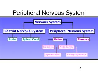
Nervous system Chapter 18 peripheral nervous system • The Spinal Nerves • The Cranial Nerves • The Autonomic Nervous System • motor nerve • visceral sensory nerve • referred pain • the innervation of some important organs
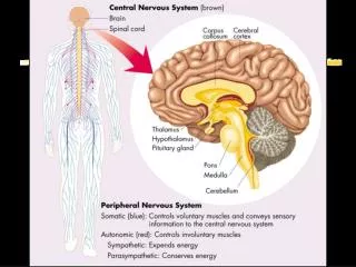
The Autonomic Nervous System central part periphery include Distribute on internal organs, blood vessels,smooth muscle and glands。 sympathetic nerve Visceral motor nerve parasympathetic nerve periphery Visceral sensory nerve Visceral motor nerveis also called autonomic nervous system or vegetative nervous system.
The composition of autonomic nervous system can be summarized as following: periphery Visceral nervous system Spinal cord Brain stem central part diencephalons cerebrum
Sympathetic nerve Visceral motor nerve Cranium part periphery Parasympathetic nerve pars sacralis Visceral sensory nerve Spinal ganglion cerebral ganglion
Visceral motor nerve The differentiation between visceral motor nerve and somatic motor nerve:
The visceral motor nerve can be divided into sympathetic nerve and parasympathetic nerve. sympathetic nerve The general view of sympathetic nerve sympathetic nerve lower centra:the T1~L3 intermediolateral nucleus paravertebral ganglia (ganglia of sympathetic trunk) interganglionic branches cervical part 3-4 ones thoracic part 10-12 ones pars lumbalis 4 ones pars sacralis 2-3 ones the two sides of pars terminalis synthetize an odd knob
prevertebral ganglia celiac ganglia superior mesenteric ganglion inferior mesenteric ganglia aorticorenal ganglia middle cervical ganglion superior cervical ganglion cervicothoracic ganglion sympathetic trunk cardiac plexus pulmonary plexus thoracic ganglia stomach superior mesenteric ganglion celiac ganglia small intestine superior mesenteric ganglion lumbar ganglion Lumbar splanchnic nerves sacral ganglia
communicating branches white communicating branches gray communicating branches spinal ganglia gray communicating branch posterior root abdominal cavity ganglion anterior root white communicating branch ganglia of sympathetic trunk
The 3 directions of preganglionic fibers of sympathetic nerve: intermediolateral nucleus—anterior root—trunk of spinal nerve—white communicating branches—sympathetic trunk ①End at the correspondent paravertebral ganglia,and change neuron. ②After trading upward or descending, terminates at the paravertebral ganglia. (T1~T6)The preganglionic fibers of intermediolateral nucleus go up to cervical part in the sympathetic trunk, and change neuron in the paravertebral ganglia of cervical part. (T6~T10)upgrading or descending in the sympathetic trunk, and change neuron in thoracic sympathetic ganglion. (T11~L3)descending in the sympathetic trunk and change neuron in the sympathetic ganglion of lumbosacral area. ③After going through paravertebral ganglia, neuron is changed inprevertebral ganglia.
The three directions of postganglionic fibre of sympathetic nerve ①The postganglionic fibre derived from sympathetic trunk return to spinal nerve via gray communicating branch, and distribute to pate, trunk and acral blood vessel, sweat glangds and arrector muscle.There aregray communicating branches between spinal nerves of 31 pairs and sympathetic trunk.The branches of spinal nerves usually contain postganglionic fibre of sympathetic nerve. ②To hold on to arteries and form correspondent nerve plexus on tunica adventitia of artery, anddistribute to the dominant organswith artery. ③Distribute directly to dominant organs from sympathetic ganglia.
superior cervical ganglion The distribution of sympathetic nerve middle cervical ganglion cervical part: superior cervical ganglion middle cervical ganglion inferior cervical ganglion the 1stinferior cervical ganglion cervicothoracic ganglion cervicothoracic ganglion cardiac plexus sympathetic trunk pulmonary plexus thoracic ganglia The distribution of postganglionic nerve fibers emerged from cervical part ganglia of sympathetic trunk: ①distribute on blood vessels, sweat glands and arrector muscles of head, neck and upper extremity. ②distribute directly to the adjacent arteries and form internal carotid plexus,external carotid plexus,subclavian plexusand vertebral plexus. ③the emergent pharyngeal branch composed of pharyngeal plexus with the pharyngeal branch of vagus nerve and cranial nerve. ④the 3 pairs of cervical ganglia of sympathetic trunk send out superior, middle and inferior cardiac nerve respectively and descend to thoracic cavity and add to cardiac plexus.
thoracic part: thoracic sympathetic nerve lie at the anterior aspect of capitulum costae thoracic ganglia 10~12 ones(the 11 ones is mostly common) branches: ①distribute on blood vessels, sweat glands and arrector muscle of thoracic and abdominal wall with 12 pairs of thoracic nerves via gray communicating branch. ②the 1st to 5th thoracic sympathetic ganglion→thoracic aortic plexus, esophageal plexus, pulmonary plexus and cardiac plexus and so on. ③the preganglionic fibre walking through the 6th to 9th thoracic ganglia constitute greater splanchnic nerve→celiac ganglia ④the preganglionic fibre walking through the 10th to 12th thoracic ganglia, constitute lesser splanchnic nerve→aorticorenal ganglia ⑤least splanchnic nerve
middle cervical ganglion superior cervical ganglion cervicothoracic ganglion sympathetic trunk cardiac plexus pulmonary plexus thoracic ganglia stomach superior mesenteric ganglion celiac ganglia small intestine superior mesenteric ganglion lumbar ganglion splanchnic nerves sacral ganglia
lumbar part 4 pairs of lumbar ganglion distribute branches thoracic ganglia There are 2 to 3 pairs of sacral ganglia and acoccygeal ganglion distribute branches: ①distribute with sacral and coccygeal nerve via gray communicating branches. ②some small branches add to pelvis plexus→pelvic cavity organs stomach celiac ganglia superior mesenteric ganglion small intestinal superior mesenteric ganglion lumbar ganglion lumbar splanchnic nerves sacral ganglia pelvic part ① Distribute to 5 pairs of lumbar nerve via gray commmunicating branches. ② The preganglionic fibre walking through lumbar ganglion — lumbar splanchnic nerves→superior and inferior mesenteric ganglion,postganglionic fibre→the digestive canal below left flexure of colon and pelvic cavity organs.
conclusions: 1、The preganglionic fibre derived from the 1st to 5th of thoracic part intermediolateral nucleus change neuron, then the postganglionic fibre arrange blood vessels, sweat glands and arrector muscle of head, neck, thoracic cavity organs and upper limb. 2、The preganglionic fibre derived from the 6th to 12th of thoracic part intermediolateral nucleus change neuron, then the postganglionic fibre arrange liver, spleen, kidney and the intestine canal above left colic flexure . 3、The preganglionic fibers derived from the 1st to 3rd of lumbar part intermediolateral nucleus change neuron, then the postganglionic fibre arrange the intestine canal and pelvic cavity organs below left colic flexure and the blood vessels, sweat glands and arrector muscle of lower limb.
parasympathetic nerve Lower center Cranial part: nucleus of oculomotor nerve superior salivatory nucleus inferior salivatory nucleus dorsal nucleus of vagus nerve Pars sacralis: the second to 4th sacral parasympathetic nucleus Parasympathetic ganglion • Head:ciliary ganglion、pterygopalatine ganglion、 • submandibular ganglion、auricular ganglion • The ganglia lie at cardiac plexus, pulmonary plexus, vesical plexus and uterovaginal plexus,and the ganglia lie at bronchus and the wall of digestive canal. The parasympathetic neuron belong to cholinergic neuron, and most of them contain VIP and CGRP and so on.
parasympathetic nerve of cranial part accessory nucleus of oculomotor nerve cranial nerve ciliary muscle contractor pupillae muscle ciliary ganglion chorda tympani submandibular gland、sublingual gland cranial nerve submandibular ganglion Superior salivatory nucleus great superficial petrosal nerve pterygopalatine ganglion lachrymal gland lesser petrosal nerve cranial nerve external salivary gland inferior salivatory nucleus auricular ganglion
dorsal nucleus of vagus cranial nerve Paraganglion of organ intramural ganglion thoracic cavity organs abdominal cavity organs (the intestine canal above left colic flexure)
parasympathetic nerve of pars sacralis sacral parasympathetic nucleus pelvic splanchnic nerves intestine canal above Left colic flexure and pelvic cavity organs paraganglion of organs intramural ganglion
the difference between sympathetic nerve and parasympathetic nervous
splanchnic plexus cardiac plexus pulmonary plexus
celiac plexus abdominal aortic plexus hypogastric plexus superior hypogastric plexus inferior hypogastric plexus (pelvic plexus)
visceral sensory nerve peripheral process geniculate ganglion inferior jugular ganglion inferior vagal ganglion ————cranial nerve —ossopharyngeal nerve —vagus nerve central process • solitary • tract nucleus →internal organ peripheral process • spinal cord(intermediomedial nucleus) central process ———sympathetic nerve —parasympathetic nerve of pars sacralis spinal ganglia →internal organ feature: 1. the pain threshold is higher. 2. dispersed visceralgia,the position isn’t exact.
referred pain When some internal organs had pathological changes, hyperaesthesis or sense of pain usually emerged on some region of body surface. This phenomenon is called referred pain. dermatomic area afferent fibers of skin (thoracic segments1~5) spinothalamic tract thoracic segments1~5 substantia gelatinosa visceral afferent fiber(thoracic segments1~5)
the innervation of some critical organ eyeball sensory nerve general sensation→nervi ciliaris longi→nasociliary nerve→ophthalmic nerve→trigeminal nerves→brain stem→nucleus sensorius nervi trigemini sympathetic nerve spinal cordT1~T2lateral horn (preganglionic fibre)→thoracic and cervical sympathetic trunk→superior cervical ganglion (exchange neuron) → (postganglionic fibre) internal carotid plexus→cavernous plexus→ciliary ganglion→dilator muscle of pupil and blood vessels parasympathetic nerve Midbraina ccessory nucleus of oculomotor nerve(E-W nucelus)(preganglionic fibre) →oculomotornerve courser→ciliary ganglion (exchange neuron)→postganglionic fibre) short ciliary nerve→pupil sphincter muscle and ciliary muscle • The excitation of sympathetic nerve of dominating eyeball will cause corectasis and vasoconstriction of iris. • The excitation of parasympathetic nerve will lead to constriction of pupil and the contraction of ciliary muscle.
Heart sensory nerve Algesthesia fiber(walk along sympathetic nerve, exceptsuperior cervical cardiac nerve)→spinal cordT1~T4,T 5 segment sensory fiber related to cardiac reflect(walk along vagus nerve) →enter into brain stem sympathetic nerve the lateral cord of spinal cord T1~T4,T 5 segment(preganglionic fiber)→the superior, middle and inferior ganglion ofsympathetic trunk, and the upper part of thoracic ganglia(exchange neuron)→give out from ganglion(the superior, middle and inferior cervical cardiac nerve and thoracic cardiac branches)→the posterior aspect and inferior aspect ofaortic arch(with the parasympathetic nerve come from vagus nerve)→cardiac plexus→heart parasympathetic nerve dorsal nucleus of vagus and ambiguous nucleus(preganglionic fibre)→walk along the rami cardiaci of vagus →cardiac ganglia(exchange neuron)→heart • The stimulation of sympathetic nerve dominating heart can induce tachycardia and the relaxation of coronary vessels. • The stimulation of vagus can induce bradycardia and the contraction of coronary vessels.