Download
1 / 15
170 likes | 213 Vues
Methods: Protein-Protein Interactions. Biochemistry 4000 Dr. Ute Kothe. k 1. P + L P-L. k -1. Equilibrium dissociation constant K D : concentration of 50% binding. [P] [L] k -1 K D = = [PL] k 1. Fraction bound (F):

E N D
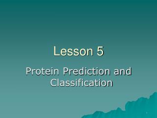
Methods:Protein-Protein Interactions Biochemistry 4000 Dr. Ute Kothe
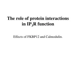
k1 P + L P-L k-1 Equilibrium dissociation constant KD: concentration of 50% binding [P] [L] k-1 KD = = [PL] k1 Fraction bound (F): [PL] [L] F = = [Ptotal] [L] + KD Remember: PC of binding
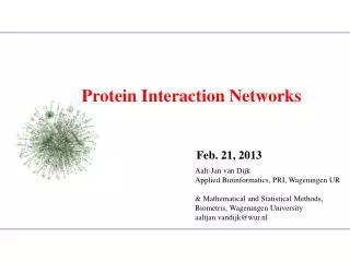
Electrophoretic mobility shift assay • Also band shift • or gel retention/retardation assay • Detection of • nucleic acid – protein interaction • protein – protein interactions • Pre- Incubation of biomolecules to form complex • Native PAGE allowing the interactions to be maintained during electrophoresis Coomassie Autoradiography truncated protein DNA Full-length protein
Electrophoretic mobility shift assay Analysis: • Coomassie stain • Ethidium bromide stain of nucleic acid • Autoradiography to detect radioactivley labelled proteins or nucleic acids • Shift of bands relative to free components indicates interaction Advantages: • qualitatively detection of interaction • low cost: no special equipment needed, low amounts of biomolecules Disadvantages: • No quantitative data (in the best rough estimation of KD) • Interacting biomolecules must have different electrophoretic mobilities
Light Scattering • Similar to Scattering of X-rays from a crystal • But: UV or VIS light, i.e. wavelength is larger than size of biomolecule • No determination of structure, but of size (molecular weight) • Size reflects formation of oligomers or complex with other protein Instrument: Compare intensity of incident light with light scattered at a given angle θ Solutions must be well filtered to avoid scattering from large dust particles! Generates monochromatic beam
Light Scattering - Principle • Principle: • Simple example: monochromatic, linearly polarized light interacting with a single molecule • The electric field of the light oscillates at the point of the single molecule • causes the molecule to have an oscillating dipole • oscillating dipole acts as mini-antenna dispersing some energy in directions other than the direction of the incident radiation = elastic Rayleigh SCATTERING
Static Light Scattering Scattering is dependent on • l, θ, and concentration – can be chosen • refractive index – measure for • polarizability in visible region of • light, can be measured • the molecular weight MW. • Determination of molecular Weight MW of a monomner (A) by measuring at various concentrations and extrapolating to c=0 (to account for solution nonideality). • What about Dimers (B)?
Dynamic Light Scattering • Instead of measuring the average light scattering in a large volume, a small volume is observed. • Fluctuations in local concentrations over time become significant • reflect diffusion of molecules • can be used to determine diffusion coefficient D
Surface Plasmon Resonance (SPR) • Sensor chip with gold film: • carries Protein 1 • Protein 2 (interaction partner) is introduced in flow channel (constant flow) • Binding interaction changes mass at surface of chip • Refractive index of chip changes • reflection angle and intensity of polarized light changes
SPR: Principle Principle: • Total reflection occurs at the critical angle which depends on the refractive index of the surface. • Energy carried by photons can be transferred to electrons in a metal at a certain wavelength (resonance) • At the resonance wavelength, almost all light is absorbed. • This creates a plasmon, a group of excited electrons in the metal surface which behave like a single electrical entity. • The plasmon generates an electrical field about 100 nm above and below the surface, called evanescent wave. Characteristic used to measure binding: • Change in chemical composition of environment of plasmon field causes a change in refractive index and thus in the resonance wavelength / in the critical angle for total reflection. • Change in mass of complex bound on surface is proportional to change in angle of totally reflected polarized light.
SPR: Results Typical Sensogram • dissociation constant (KD) from signal intensity in dependence of ligand concentration • apparent association and dissociation constants (kon, koff) from signal change during injection of ligand / removal of ligand • Disadvantage: • No equilibrium method • Constant flow of ligand
Isothermal Titration Calorimetry (ITC) • Determine absorption or release of heat (q) upon binding of a ligand to a biomolecule • heat is proportional to enthalpy DH°(T) and number of moles complex (nPL = V * [PL]): q = DH°(T) * V * [PL] [L] q = DH°(T) * V * [Ptotal] [L] + KD • By measuring q at various ligand concentrations while knowing the volume and total protein concentration, DH°(T) and KD can be determined! Remember: DG°(T) = - RT ln KA and DG°(T) = DH°(T) – T*DS°(T) • DG°(T) and DS°(T) can be determined!
ITC - Instrument • stepwise addition of ligand into protein • solution of known concentration • by comparison with a reference cell • containing only buffer, the energy is • measured which is required to maintain • a constant termperature over time • heat q is obtained by integrating peak • area over time • Disadvantage: • Significant amounts of proteins needed • (Size of cell 1 – 2 ml) • Tight binding interactions can not be studied (KD should be in µM range)
ITC - Data • Binding between core binding domain of exterior glyco-protein gp120 form the HIV-1 virus and the CD4 receptor fo the target host cell • DH° = -263 kJ/mol, • KA = 5 x 10⁶ M-1
Other Methods • Fluorescence (FRET) • Size Exclusion Chromatography • Immuno precipitation • Affinity chromatography • Crosslinking • Analytic ultracentrifugation • Mass spectrometry