Download
1 / 1
10 likes | 166 Vues
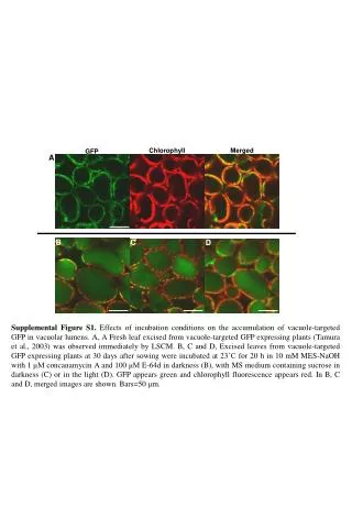
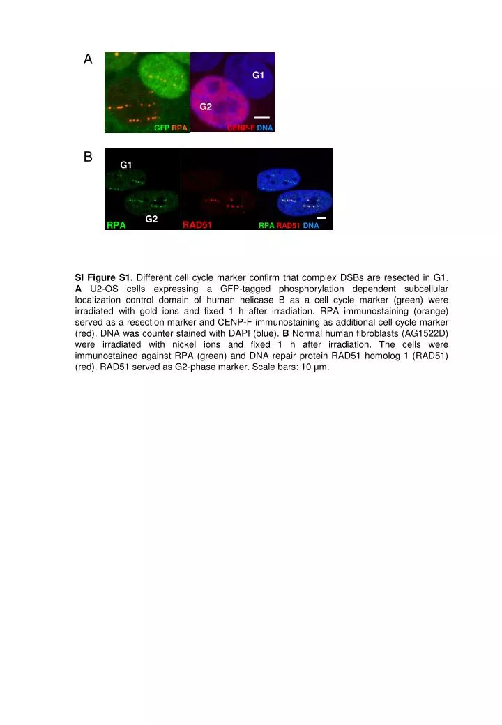
G1. G2. GFP RPA. CENP-F DNA. G1. c. G2. RAD51. RPA. RPA RAD51 DNA. A. B.

E N D
G1 G2 GFP RPA CENP-FDNA G1 c G2 RAD51 RPA RPA RAD51DNA A B • SI Figure S1. Different cell cycle marker confirm that complex DSBs are resected in G1. AU2-OS cells expressing a GFP-tagged phosphorylation dependent subcellular localization control domain of human helicase B as a cell cycle marker (green) were irradiated with gold ions and fixed 1 h after irradiation. RPA immunostaining (orange) served as a resection marker and CENP-F immunostaining as additional cell cycle marker (red). DNA was counter stained with DAPI (blue). B Normal human fibroblasts (AG1522D) were irradiated with nickel ions and fixed 1 h after irradiation. The cells were immunostained against RPA (green) and DNA repair protein RAD51 homolog 1 (RAD51) (red). RAD51 served as G2-phase marker. Scale bars: 10 μm.