Download
1 / 53
840 likes | 1.64k Vues
Wound Care and Dressing. Presented By Dr. Osama Kentab, MD, FAAP, FACEP Assistant Professor of Pediatrics and emergency Medicine King Saud Bin Abdulaziz University for Health sinces Riyadh. THE SKIN. FUNCTIONS OF THE SKIN. Regulates body temperature.

E N D
Wound Care and Dressing Presented By Dr. Osama Kentab, MD, FAAP, FACEP Assistant Professor of Pediatrics and emergency Medicine King Saud Bin Abdulaziz University for Health sinces Riyadh
FUNCTIONS OF THE SKIN • Regulates body temperature. • Prevents loss of essential body fluids, and penetration of toxic substances. • Protection of the body from harmful effects of the sun and radiation. • Excretes toxic substances with sweat ( waste removal). • Mechanical support. • Immunological function mediated by Langerhans cells. • Sensory organ for touch, heat, cold, socio-sexual and emotional sensations. • Vitamin D synthesis from its precursors under the effect of sunlight and introversion of steroids.
Wound-definitions(Manley, Bellman, 2000) • A loss of continuity of the skin or mucous membrane which may involve soft tissues, muscles, bone and other anatomical structure. • Any disruption to layers of the skin and underlying tissues due to multiple causes including trauma, surgery, or a specific disease state.
WOUND HEALING Classification of wound healing (According to the amount of tissue loss) • Primary intention healing • Secondary intention healing • Tertiary intention healing
PHASES OF WOUND HEALING Healing is a quality of living tissue; it is also referred to as regeneration (renewal) of tissue. • The inflammatory phase (3-6 days) • The regenerative (Proliferative) phase (day 4-day21) • The maturation (Remodeling) phase (day 21- 1 or 2 yrs) (Manley, Bellman, 2000)
The inflammatory phase (Initiated immediately after injury and last 3-6 days Injury /damage Cells Histamine Blood Clot Dry Vasodilation Permeability Uniting the wound edges Neutrophils& Monocytes • -Dilated blood vessels • Microcirculation slow down Oedema& Engorgement 0-3 days
The Regenerative (Proliferative) phase Blood vessels near the edge of the wound become porous Begins 2-3 days of injury Lasting up to 2-3 weeks Allowing excess moisture to escape - Resultant tissue filling is referred To as granulation tissue - process of wound contraction begins Macrophage activity Traps other blood cells & damaged blood vessels Begin to regenerate within the wound margins Stimulates Formation& multiplication of fibroblasts This fibrous network Which - Laying down of a ground substance - Beginning the synthesis of collagen fibers (granulation tissue ) migrate along fibrin threads Resulting
The Maturative phase • Begins about day 21 and can extend up to 6 months up to one or two years after the injury. • Fibroblasts continue to synthesize collagen • The collagen fibers recognized into a more orderly structure • The scar become a thin ,less elastic, white line
Factors affecting wound healing • Developmental consideration/Age • Nutrition • Life-style • Medication • Infection • Wound perfusion
Classification of surgical wounds according to the degree of contamination Clean wounds: Operations in which a viscus is not opened. This category includes non- traumatic, uninfected wounds where is no inflammation encountered and no break in technique has occurred. Clean-contaminated: A viscus is entered but without spillage of contents. This category included non- traumatic wounds where a minor break in technique has occurred.
Classification of surgical wounds cont’d(Altmeire 1997, Ayliffe & Lowbury 1992, NAS 1996) Contaminated: Gross spillage has occurred or a fresh traumaticwound from a relatively clean source. Acute non-purulent inflammation may also be encountered. Dirty or infected : Old traumatic wounds from a dirty source, with delayed treatment, devitalised tissue, clinical infection, faecal contamination or a foreign body.
Classification of wounds by depth • Partial-thickness: Confined to the skin, the dermis and epidermis. • Full-thickness : Involve the dermis, epidermis, subcutaneous tissue, and possibly muscle and bone Partial Thickness Full Thickness
Wound assessment cont’d(Hahn,Olsen,Tomaselli, Goldberg ,2004) What to assess? • Location • Dimensions/Size • Tissue viability • Exudate/Drainage • Periwound condition • Pain • Stage or extent of tissue damage , dictates how often a wound is reassessed • Swelling
Diagnoses • Risk for Impaired Skin Integrity • Impaired Skin Integrity • Impaired Tissue Integrity • Risk for Infection • Pain
Risk Factors Which Increase Patient Susceptibility to infection(Manley.K, Bellman. L,2000) A- Intrinsic risk factors: • Extremes age: Defined as “ Children aged 1 year and under, and people aged 65 years and over’. • Underling Conditions/Disorders • Diabetes • Respiratory disorders • Blood disorders • Smoking • Nutrition and build
Risk Factors Which Increase Patient Susceptibility to infection cont’d(Manley.K, Bellman. L,2000) B- Extrinsic risk factors: • Drug therapy as a risk factor: e.g. Cytotoxic drugs • Break in the integrity of the skin • Items such as foreign bodies • Bypassing of defense mechanisms through devices e.g. Intubations
S&S of Presence of Infection • Wound is swollen. • Wound is deep red in color. • Wound feels hot on palpation. • Drainage is increased and possibly purulent. • Foul odor may be noted. • Wound edges may be separated with dehiscence present.
Types of Wound Drainage Exudateis material, such as fluid and cells, that has escaped from blood vessels during the inflammatory process and deposited in or on tissue surfaces. The Nature and amount of exudate vary according to: Tissue involved, Intensity and duration of the inflammation, and the presence of microorganisms. 1. Serous Exudate • Mostly serum • Watery, clear of cells • E.g., fluid in a blister
A purulent Exudate • Is thicker than serous exudate because of the presence of pus. • It consists of leukocytes, liquefied dead tissue debris, dead and living bacteria. • The Process of pus formation is referred to as suppuration, and the bacteria that produce pus are called pyogenic bacteria. • Purulent exudate vary in color, some acquiring tinges of blue, green, or yellow. The color may depend on the causative organism.
A sanguineous (hemorrhagic) Exudate • It consists of large amount or blood cells, indicating damage to capillaries that is very severe enough to allow the escape of RBCs from plasma • This type of exudate is frequently seen in open wounds. • we often need to distinguish whether the exudate is dark or bright. Bright indicate fresh blood, whereas dark exudate denotes older bleeding.
Complications of Wounds • Infection • Hemorrhage • Dehiscence and possible evisceration • Fistula formation
The RYB color code(Stotts,1999) • This concept is based on the color of the open wound rather than the depth or size of the wound. • On this scheme, the goal of wound care is to protect ( cover) red, cleanse yellow, and debride black. • The RYB code can be applied to any wound allowed to heal by secondary intention. R=Red Y=Yellow B= Black
Red wounds • Usually in the late regeneration phase of tissue repair (ie, developing granulation tissue) and are clean and uniformly pink in appearance • They need to be protected to avoid disturbance to regenerating tissue. Examples are superficial wounds, skin donor sites, and partial- thickness or second – degree burns.
How to protect red wounds: • Gentle cleansing • Avoid the use of dry gauze or wet- to-dry saline dressings. • Applying a topical antimicrobial agent. • Appling a transparent film or hydrocolloid dressing. • Changing the dressing as infrequently as possible.
Yellow wounds • Characterized primarily by liquid to semiliquid ”slough” that is often accompanied by purulent drainage. • clean yellow wounds to absorb drainage and remove nonviable tissue. Methods used may include . • Applying wet-to-wet dressing; irrigating the wound; using absorbent dressing material such as impregnated nonadherent, hydrogel dressing, or other exudate absorbers; and a topical antimicrobial to minimize bacterial growth.
Black Wound • Covered with thick necrotic tissue or Eschar. • e.g.. third degree burns and gangrenous ulcer. • Required debridement . • When the eschar is removed, the wound is treated as yellow, then red.
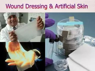
Purposes of wound dressing • To protect the wound from mechanical injuries • To protect the wound from microbial contamination • To provide or maintain high humidity of the wound • To provide thermal insulation • To absorb drainage and /or debride a wound
Purposes of wound dressing 6. To prevent hemorrhage (when applied as a pressure dressing or with elastic bandages). 7. To splint or immobilize the wound site and thereby facilitate healing and prevent injury. 8. To provide psychological (aesthetic) comfort.
Principles of asepsis The aim: • Guarantee the safety of the equipment used (cleaning/disinfection/sterilisation). • Reduce the level of microbial contamination of the site requiring manipulation (antisepsis). • Ensure that no microorganisms are introduced (asepsis).
Cleaning : Is the removal of dirt, debris and organic material. Disinfection: Removes or destroys harmful microorganisms but not bacterial spores or slow viruses. Sterilization: is the complete destruction or removal of all living microorganisms including bacterial spores. Antisepsis: is the reduction of the number of microorganisms already present on the body site prior to a procedure. Asepsis: Procedure designed to prevent any introduction of microorganisms to the site achieved by anon-touching technique and use of sterile gloves
Evaluation of Wounds • ABC’s first Always! • Ensure hemostasis • Saline gauze dressing • Compression • Remove obstructions • Rings, clothing, other jewelry • History
Tetanus status Allergies Medications Comorbidities Previous scar formation History • Symptoms • Type of Force • Contamination • Event • Potential for foreign body • Function • Non-accidental trauma
Vascular function Tendon function Underlying structures Wound contamination Foreign bodies Wound Examination • Location • Size • Shape • Margins • Depth • Alignment with skin lines • Neuro function
Wound Consultation • Tarsal plate or lacrimal duct • Open fracture or joint space • Extensive facial wounds • Associated with amputation • Associated with loss of function • Involves tendons, nerves, or vessels • Involves significant loss of epidermis • Any wound that you are uncertain about
Wound Preparation - Hemostasis • Physical vs. chemical • Direct pressure • Epinephrine • Gelfoam • Cautery • Refractory • Use a tourniquet
Visual inspection Imaging Glass, metal, gravel fragments >1mm should be visible on plain radiographs Organic substances and plastics are usually radiolucent Always discuss and document possibility of retained foreign body Wound Preparation – Foreign Body Removal
Wound Preparation – Irrigation • Local anesthesia prior to irrigation • Do not soak the wound • Use normal saline • Large syringe (60mL) with Zerowet attachment • Do not use iodine, chlorhexidine, peroxide or detergents
Wound Preparation – Debridement • Removes foreign matter & devitalized tissue • Creates sharp wound edge • Excision with elliptical shape • Respect skin lines
Wound Preparation – Antibiotics • Infections occur in ~3-5% of traumatic wounds seen in the ED • Factors that increase risk • Heavily contaminated wound, especially with soil • Immunocompromised patients • Diabetics • Human bites > animal bites • Most important prevention adequate irrigation & debridement
Wound Preparation – Antibiotics • Dog & cat bites • Cover pasteurella • Augmentin • Human bites • Cover eikenella • Augmentin • Puncture wounds • Cover pseudomonas • Cipro, levaquin
Wound Preparation – Tetanus Prophylaxis • Clean wounds • Incomplete immunization toxoid • >10 years, then give toxoid • Tetanus prone wound • Incomplete immunization • Toxoid & immune globulin • > 5 years, give toxoid • Remember to think about rabies!
Guidelines for cleaning wounds • Use physiologic solution, such as isotonic saline or lactated ringer solution. • When possible , warm the solution to body temperature before use. • If the wound is grossly contaminated by foreign material , bacteria, slough, or necrotic tissue clean the wound at every dressing change. • If a wound is clean , has little exudate , and reveals healthy granulation tissue , avoid repeated cleaning.
Use gauze squares . • Consider cleaning superficial noninfected wound by irrigating them with normal saline rather than using mechanical means. • To retain wound moisture , avoid drying a wound after cleaning it.
Ideal Dressing • provide mechanical protection • protect against secondary infection • non adherent and easily removed without trauma • leave no foreign particles in the wound • remove excess exudates • cost effective • offer effective pain relief.
Burns: First Contact Assessment site depth surface area involved age of patient other influencing factors
Superficial Burn Characteristics • epidermis only • erythema (vasodilatation) • tenderness (nerve irritability) • oedema.
Superficial Partial Burn Characteristics • epidermis and outer dermis • blisters (fluid shift) • shedding of skin • painful exposed (nerve endings to kinins) • bleeds when pricked with needle • hair present (hard to pull out) • full sensation • blanches on pressure.
Burn Surface Area • Wallace’s rule of nines • Lund and Browder chart • closed palmar hand of victim = 1% of body surface area.