Download

1 / 47
470 likes | 558 Vues
Histopathology and staging of breast cancer Prof T J Stephenson. Physiological conditions. Pregnancy and lactation Gynaecomastia Involution. Fibrocystic change. Adenosis Fibrosis Cysts Epithelial hyperplasia (+/- atypia) Apocrine metaplasia. Inflammatory conditions. Acute mastitis

E N D
Histopathology and staging of breast cancer Prof T J Stephenson
Physiological conditions • Pregnancy and lactation • Gynaecomastia • Involution
Fibrocystic change Adenosis Fibrosis Cysts Epithelial hyperplasia (+/- atypia) Apocrine metaplasia
Inflammatory conditions • Acute mastitis • Abscess • Duct ectasia • Fat necrosis
Benign tumours • Fibroadenoma • Adenoma • Intraduct papilloma • Connective tissue neoplasms
Fibroadenoma Commonest benign tumour Biphasic Very characteristic clinico-pathological features
← Circumscribed lesion Epithelium and stroma →
Intraduct papilloma • Middle aged women • Blood stained nipple discharge from large ducts • Typical papillary structures with fibrovascular cores
Lobular carcinoma in situ • 6% of breast malignancies • Premenopausal • Impalpable • Up to ⅓ develop invasive cancer if only biopsied • Equal risk in both breasts, unless PLCIS which behaves like DCIS
Ductal carcinoma in situ • Mass / Paget’s / discharge / screening • Different histological types / grades • All have slightly different characteristics
Risk factors • Female gender • Cancer in other breast • Long interval between menarche and menopause • Age at first full-term pregnancy • Not breast feeding • Obesity and high fat diet • Family history • Geographical factors • Histological risk factors
Breast cancer 20% of all cancers in women, (second only to lung) Commonest cancer in the UK Commonest cause of death in women 35 – 55 In UK, women have 1:8 lifetime chance of developing it (but 1:33 by 50) 48417 new cases per year (2009) and rising 11556 deaths in UK per year (2010) and falling
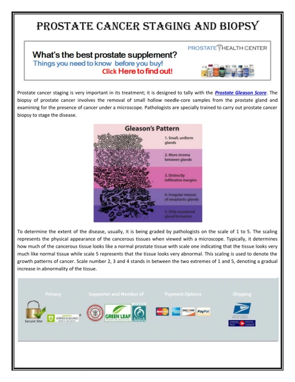
Elston/Ellis modification of Bloom and Richardson Grading Detailed Criteria used in Histologic Grade Glandular (Acinar)/Tubular Differentiation Score 1: >75% of tumor area forming glandular/tubular structures Score 2: 10% to 75% of tumor area forming glandular/tubular structures Score 3: <10% of tumor area forming glandular/tubular structures Nuclear Pleomorphism Score 1: Nuclei small with little increase in size in comparison with normal breast epithelial cells, regular outlines, uniform nuclear chromatin, little variation in size Score 2: Cells larger than normal with open vesicular nuclei, visible nucleoli, and moderate variability in both size and shape Score 3: Vesicular nuclei, often with prominent nucleoli, exhibiting marked variation in size and shape, occasionally with very large and bizarre forms Mitotic Count The mitotic count score criteria vary depending on the field diameter of the microscope used by the pathologist. The pathologist will count how many mitotic figures are seen in 10 high power fields. Using a high power field diameter of 0.50 mm, the criteria is as follows: Score 1: less than or equal to 7 mitoses per high power field Score 2: 8-14 mitoses per high power field Score 3: equal to or greater than 15 mitoses per high power field Overall Grade Grade 1: scores of 3, 4, or 5 Grade 2: scores of 6 or 7 Grade 3: scores of 8 or 9
Spread and Staging • Local • Intra-cavity • Lymphatic • Blood stream
TNM definitions • Primary tumor (T): • TX: Primary tumor cannot be assessed T0: No evidence of primary tumor Tis: Carcinoma in situ; intraductal carcinoma, lobular carcinoma in situ, or • Paget's disease of the nipple with no associated tumor. Note: Paget's disease associated with a tumor is classified according to the size of the tumor. • T1: Tumor 2.0 cm or less in greatest dimension • T1mic: Microinvasion 0.1 cm or less in greatest dimension T1a: Tumor more than 0.1 but not more than 0.5 cm in greatest dimension T1b: Tumor more than 0.5 cm but not more than 1.0 cm in greatest dimension T1c: Tumor more than 1.0 cm but not more than 2.0 cm in greatest dimension • T2: Tumor more than 2.0 cm but not more than 5.0 cm in greatest dimension • T3: Tumor more than 5.0 cm in greatest dimension • T4: Tumor of any size with direct extension to (a) chest wall or (b) skin, • only as described below. Note: Chest wall includes ribs, intercostal muscles, and serratus anterior muscle but not pectoral muscle. T4a: Extension to chest wall T4b: Edema (including peau d'orange) or ulceration of the skin of the • breast or satellite skin nodules confined to the same breast T4c: Both of the above (T4a and T4b) T4d: Inflammatory carcinoma*
TNM definitions • Regional lymph nodes (N): • NX: Regional lymph nodes cannot be assessed (e.g., previously removed) N0: No regional lymph node metastasis N1: Metastasis to movable ipsilateral axillary lymph node(s) N2: Metastasis to ipsilateral axillary lymph node(s) fixed to each other or • to other structures N3: Metastasis to ipsilateral internal mammary lymph node(s) Pathologic classification (pN): • pNX: Regional lymph nodes cannot be assessed (not removed for pathologic • study or previously removed) pN0: No regional lymph node metastasis pN1: Metastasis to movable ipsilateral axillary lymph node(s) • pN1a: Only micrometastasis (none larger than 0.2 cm) pN1b: Metastasis to lymph node(s), any larger than 0.2 cm • pN1bi: Metastasis in 1 to 3 lymph nodes, any more than 0.2 cm and all • less than 2.0 cm in greatest dimension pN1bii: Metastasis to 4 or more lymph nodes, any more than 0.2 cm and • all less than 2.0 cm in greatest dimension pN1biii: Extension of tumor beyond the capsule of a lymph node • metastasis less than 2.0 cm in greatest dimension pN1biv: Metastasis to a lymph node 2.0 cm or more in greatest dimension pN2: Metastasis to ipsilateral axillary lymph node(s) fixed to each other • or to other structures pN3: Metastasis to ipsilateral internal mammary lymph node(s)
TNM definitions • Distant metastasis (M): • MX: Presence of distant metastasis cannot be assessed M0: No distant metastasis M1: Distant metastasis present (includes metastasis to ipsilateral • supraclavicular lymph nodes)
Prognosis • PI = 0.2(cm size) + grade (1, 2 or 3) + LN stage* (1, 2 or 3) • LN stage: 1 = not involved 2 = 1 – 3 nodes involved 3 = 4 or more nodes involved or level 3