Download
1 / 11
170 likes | 599 Vues
Fig 2.1A: Axial Flair MRI. Fig 2.1B: Axial T1 Weighted (Wtd.) MRI. Fig 2.1C: Post-Contrast Axial T1 Wtd. MRI. 56 Year-old male with 3 month history of gradual weakness of the right upper extremity. Diagnosis: ???. Fig 2.1D: Post Contrast Coronal T1 Wtd. MRI.

E N D
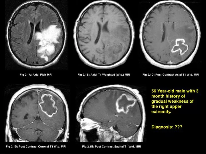
Fig 2.1A: Axial Flair MRI Fig 2.1B: Axial T1 Weighted (Wtd.) MRI Fig 2.1C: Post-Contrast Axial T1 Wtd. MRI 56 Year-old male with 3 month history of gradual weakness of the right upper extremity. Diagnosis: ??? Fig 2.1D: Post Contrast Coronal T1 Wtd. MRI Fig 2.1E: Post Contrast Sagital T1 Wtd. MRI
Fig 2.2A: Axial Flair MRI Fig 2.2B: Axial T1 Wtd. MRI Fig 2.2C: Post-Contrast Axial T1 Wtd. MRI 51 Year-old male with history of seizures and was involved in a car accident, leading to imaging studies. Diagnosis: ??? Fig 2.2D: Post Contrast Coronal T1 Wtd. MRI
Fig 2.3A: Axial Flair MRI Fig 2.3B: Axial T1 Wtd. MRI Fig 2.3C: Post-Contrast Axial T1 Wtd. MRI 19 Year-old young lady with seizures and headache Diagnosis: ??? Fig 2.3D: Post Contrast Sagital T1 Wtd. MRI
Fig 2.4A: Axial T2 Wtd. MRI Fig 2.4B: Axial T1 Wtd. MRI Fig 2.4C: Post-Contrast Axial T1 Wtd. MRI 12 Year-old boy with headache, nausea and vomiting for three months. Diagnosis: ??? Fig 2.4D: Post Contrast Coronal T1 Wtd. MRI
Fig 2.5A: Axial Flair MRI Fig 2.5B: Axial T1 Wtd. MRI Fig 2.5C: Post-Contrast Axial T1 Wtd. MRI 60 Year-old male with six month history of gradual memory loss, disorientation and headache. Diagnosis: ??? Fig 2.5D: Post Contrast Coronal T1 Wtd. MRI
Match the following Gliomas shown in figs 2.1 thru 2.5 to WHO (World Health Organization) Classification: Fig. 2.1 A. Butterfuly Glioblastoma Fig. 2.2 B. Grade 2 Astrocytoma Fig. 2.3 C. Pilocytic Astrocytoma (Grade 1 Astrocytoma) Fig. 2.4 D. Glioblastoma (Grade 4 Astrocytoma) See Next Panel for Correct Answers Fig. 2.5 E. Anaplastic Astrocytoma Grade 3
Correct Answers Match the following Gliomas shown in figs 2.1 thru 2.5 to WHO (World Health Organization) Classification: Fig. 2.1 D. Glioblastoma (Grade 4 Astrocytoma) Fig. 2.2 E. Anaplastic Astrocytoma Grade 3 B. Grade 2 Astrocytoma Fig. 2.3 C. Pilocytic Astrocytoma (Grade 1 Astrocytoma) Fig. 2.4 Fig. 2.5 A. Butterfuly Glioblastoma
E Fig 2.1A: Axial Flair MRI Fig 2.1B: Axial T1 Weighted (Wtd.) MRI Fig 2.1C: Post-Contrast Axial T1 Wtd. MRI An irregular enhancing ring lesion (arrow) is seen involving the left parietal lobe. Tumor is associated with edema (E) best noticed on FLAIR image (A). DIAGNOSIS: GLIOBLASTOMA • Grade IV Astrocytoma (WHO Classification) • Older Patient • Imaging Features: Tumor with irregular peripheral enhancement with central necrosis. • Median survival is 1 – 2 years following Surgery/Radiation therapy/Chemotherapy.
Fig 2.2A: Axial Flair MRI Fig 2.2B: Axial T1 Wtd. MRI Fig 2.2C: Post-Contrast Axial T1 Wtd. MRI Fig 2.2D: Post Contrast Coronal T1 Wtd. MRI An ill-defined non-enhancing tumor (yellow arrows) is seen in the left parietal lobe with spotty areas of enhancement (red arrows). DIAGNOSIS: Anaplastic Astrocytoma • Grade III Astrocytoma (WHO Classification) • Usually seen between 40 – 60 years of age • Imaging Features: Ill-defined non-enhancing tumor with or without feeble enhancement. • Median survival is 5 – 6 years following Surgery/Radiation therapy/Chemotherapy.
Fig 2.3A: Axial Flair MRI Fig 2.3B: Axial T1 Wtd. MRI Fig 2.3C: Post-Contrast Axial T1 Wtd. MRI Non-enhancing tumor (arrow) involving the right temporal lobe. DIAGNOSIS: GRADE III ASTROCYTOMA (LOW GRADE) • Children and young adults • Imaging Features: Non-enhancing tumors. Calcification can be seen. • Total cure from surgery to median survival of 7- 10 years.
Fig 2.4A: Axial T2 Wtd. MRI Fig 2.4B: Axial T1 Wtd. MRI Fig 2.4C: Post-Contrast Axial T1 Wtd. MRI A large cystic tumor (yellow arrow) with a mural enhancing nodule (red arrow) is seen within the left cerebellar hemisphere. DIAGNOSIS: PILOCYTIC ASTROCYTOMA • Grade I Astrocytoma (WHO Classification) • Children and young adults • Imaging Features: cyst within enhancing tumor nodule. • Surgical resection can result in complete cure.