Download
1 / 17
760 likes | 3.57k Vues
Transmission Electron Microscopy (TEM). By Austin Avery. Overview. What is Transmission Electron Microscopy? History of TEM Theory of TEM The instrumentation How is TEM useful? Pros/Cons Summary. Transmission Electron Microscopy.

E N D
Transmission Electron Microscopy (TEM) By Austin Avery
Overview • What is Transmission Electron Microscopy? • History of TEM • Theory of TEM • The instrumentation • How is TEM useful? • Pros/Cons • Summary
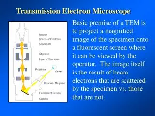
Transmission Electron Microscopy • TEM is an instrumental technique that uses a tiny focused beam of electrons. • These electrons interact with a very thin and tiny sample, usually only a couple atoms thick. • After passing through the sample the electrons have changed course slightly and are detected by a photographic film or a CCD camera.
History of TEM • In 1931 Max Knoll and Ernst Ruska developed the first TEM microscope, considered the first ‘electron microscopy’. • The group was first interested in further developing the resolution of Cathode Ray Oscilloscopes. • After WW2 Ruska was finally able to produce the first TEM with 100,000x resolution power.
TEM Theory • The reason a TEM can have such a large resolution factor at such a small size is due to the de Broglie λ of electrons. • The wavelength λe= h/√[2moE(1+(E/2moc2))] of electrons where h is planck’s constant, mo=9.11x10⁻³¹kg, E is the energy of the accelerated electron, and c is speed of light.
Theory • Because electrons have wave-particle duality properties, they can be produced at a specific energy and wavelength and then analyzed after being affected by a sample • Max resolution d=λ/(2n sin α) • Wavelegthλ • Numerical Aperture
Instrumentation • The TEM consists of an Electron gun made from a Tungsten filament or a Lanthanum hexaboride crystal • The e-gun is charged with about 100-300 kV before any electrons of reasonable energy will be released from the source. • Then a series of electromagnetic and electrostatic lenses direct the electrons into a beam
Optics • There are 3 sets of lens on a typical TEM • Condenser lens: Electron beam formation • Objector lens: focus of beam onto sample • Projector lens: expands electron beam into analytical form onto analysis screen or CCD • Magnification adjustments are made by varying the current through the quadrupole or hexapole lenses
Vacuum Components • Vacuum system: ~10-4 to 10-9 Pa • Very important to prevent arc between cathode and ground • Mean free path of electrons • Better beam focusing, less interaction with gas molecules
Sample Components • Sample grids: Usually 3mm diameter mesh ring with 1-100μm size squares made of Cu, Mb, Au, or Pt • Samples inserted into section with air locks to prevent large decreases in vacuum pressure • Stages are designed to adjust the samples orientation when inside the TEM for more accurate readings
Electron Lens Components • Electron lens: Focus parallel rays at a constant length • TEM lenses are usually electromagnetic • Made of Fe, Fe-Co, or Ni-Co because of their magnetic properties like magnetic saturation, hysteresis, and permeability
Aperture Components • Filter electrons that can stray from the beam path and affect image quality • Decrease beam intensity, helpful with beam sensitive samples • Can be fixed or moveable, depending on the quality of the instrument or manufacturer • Made of metallic substances thick enough to stop stray electrons but allow axial electrons through
Why use a TEM? • A TEM is able to form images of sample molecules and atoms 10’s of thousands times smaller than any visible light microscopes • Using the stage tuning a “tilt series” can be developed to resolve 3D images of samples
Scan Types and Uses • Bright Field • Diffraction Contrast • Dark Field Image • Crystallography-lattice defects • Biological specimen-many • Sub-atomic ratios
Pros/Cons • Pros: • Very high resolution • Requires very little sample to test • Quantitative and Qualitative • Can be modified in many ways to account for different substances and requirements • Cons: • Tough sample prep. • Hour consuming runs to get a few images • Small field of view, may take several runs to find what is being studied • Sample destruction, especially biological samples
Summary • TEM is useful for small, nanoscaleanalytes • TEM can create 3D images of samples • TEM can be modified for different types of molecules and atoms • TEM is not cheap • TEM is GOOD! • And that’s the way the cookie crumbles…
References • Kirkland, E (1998). Advanced computing in Electron Microscopy. Springer. • Hubbard, A (1995). The Handbook of surface imaging and visualization. CRC Press • Joachim Frank, editor (2006). Frank, J. ed. Electron tomography: methods for three-dimensional visualization of structures in the cell. Springer