Download
1 / 69
840 likes | 3.28k Vues
Spinal Stenosis. Niall Craig Consultant Orthopaedic Spinal Surgeon Aberdeen 25 th February 2011. Plan. Anatomy History Clinical Findings Pathophysiology Pain Sources Investigation Treatment. Spinal Stenosis. Coined 1855-60 Greek derivation Pathology – noun

E N D
Spinal Stenosis Niall Craig Consultant Orthopaedic Spinal Surgeon Aberdeen 25th February 2011
Plan • Anatomy • History • Clinical Findings • Pathophysiology • Pain Sources • Investigation • Treatment
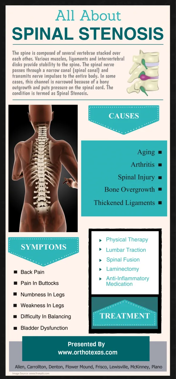
Spinal Stenosis • Coined 1855-60 • Greek derivation • Pathology – noun • Narrowing or stricture of a passage or canal • Commonest in the cervical and lumbar spine • Also occurs in the thoracic region
History HenkVerbiest First coined expression lateral stenosis in 1940s Dutch surgeon Now we think of this as lateral recess or foraminal stenosis
Spinal Canal Development 185 skeletons from the Spitalfield Collection at the Natural History Museum - French Huguenots (1646 and 1852) date of birth and death known 41 infants between 15 months and 5 years of age L1 to L4 the cross sectional area and the mid sagittal diameters - fully mature by 1 year canal size reached maturity at L5 by 5 years of age gradual trend from a circular towards a trefoil shape at L5 – trefoil shape not present prior to adulthood.
Anatomy C – central zone F – foramen E – extraforaminal zone Normal relationships Remember most people with symptoms will not have normal anatomy
Anatomy Normal canal showing space available space for thecal sac and exiting roots Specimen courtesy of Prof Rauschning Uppsala
Lateral Recess Stenosis • Lateral region is compartmentalized into entrance zone, mid zone, exit zone, and far-out stenosis
Classification • Keim et al mention the following simplistic LSS anatomical classification scheme:2 • Lateral, secondary to SAP hypertrophy • Medial, secondary to IAP hypertrophy • Central, due to hypertrophic spurring, bony projection, or ligamentum flavum/laminar thickening • Fleur de lis (clover leaf), from laminar thickening with subsequent posterolateral bulging
Lateral Recess Stenosis • Lateral recess stenosis (ie, lateral gutter stenosis, subarticular stenosis, subpedicular stenosis, foraminal canal stenosis, intervertebral foramen stenosis) - narrowing (less than 3-4 mm) between the facet superior articulating process (SAP) and posterior vertebral margin - impinge the nerve root and subsequently elicit radicular pain.
Entrance Zone • The entrance zone - medial to the pedicle and SAP – stenosis from facet joint SAP hypertrophy. • Other causes - developmentally short pedicle and facet joint morphology, as well as osteophytosis • Disc prolapse anterior to the nerve root • The lumbar nerve root compressed below SAP retains the same segmental number as the involved vertebral level (eg, L5 nerve root is impinged by L5 SAP).
Mid Zone • Mid zone extends from the medial to the lateral pedicle edge. Mid-zone stenosis arises from osteophytosis under the pars interarticularis and bursal or fibrocartilaginous hypertrophy at a spondylolytic defect
Exit Zone • Exit-zone stenosis involves an area surrounding the foramen and arises from facet joint hypertrophy and subluxation, as well as superior disc margin osteophytosis. Such stenosis may impinge the exiting spinal nerve
Extra-canalicular Stenosis • Far-out (extracanalicular) stenosis entails compression lateral to the exit zone • Occurs with far lateral vertebral body endplate osteophytosis and when the sacral ala and L5 transverse process impinge on the L5 spinal nerve
History • Patient will describe buttock pain or leg pain • Heaviness weakness • Tiredness in legs on walking • Neurogenic claudication
History • Lumbar spinal stenosis classically presents as bilateral neurogenic claudication • Unilateral radicular symptoms may result from severe foraminal or lateral recess stenosis • Patients, typically aged more than 50 years • insidious-onset NC - intermittent, crampy, diffuse radiating thigh or leg pain with associated paresthesias • Leg pain affects 90% of patients with LSS
History • Retrospective review of 75 patients with radiographically confirmed LSS, reports of weakness, numbness or tingling, radicular pain, and NC were in almost equal proportions. The most common symptom was numbness or tingling of the legs • NC pain exacerbated by standing erect and downhill ambulation, alleviated with lying supine more than prone, sitting, squatting, and lumbar flexion • Getty et al documented 80% pain diminution with sitting and 75% with forward bending.9
History • Lumbar spinal canal and lateral recess cross-sectional area increases with spinal flexion and decreases with extension • cross-sectional area is reduced 9% with extension in the normal spine and 67% with severe stenosis • Penning rule of progressive narrowing implies that the more narrowed the canal by stenosis, the more it narrows with spinal extension • Schonstrom et al have shown that spinal compressive loading from weight bearing reduces spinal canal dimensions
History • NC not exacerbated with biking, uphill ambulation, and lumbar flexion and is not alleviated with standing • compensate for symptoms by flexing forward, slowing their gait, leaning onto objects (eg, over a shopping trolley) and limiting distance of ambulation. Unfortunately this promotes disease progression and vertebral fracture. Pain radiates downward in NC and up in vascular claudication • Hall et al note the presence of radiculopathy in 6% and NC in 94% of LSS patients
History • Distinguishing between neurogenic and vascular claudication is important • Treatments and implications quite different • Vascular claudication is a manifestation of peripheral vascular disease and arteriosclerosis. Other vessels, including the coronary, vertebral, and carotid, are also often affected • Remember neurogenic and vascular claudication may occur together
Pathophysiology • Congenital • Narrow canal • Short pedicles • Arrested growth • Achondroplasia
Congenital or Primary • Congenital malformations and developmental flaws • incomplete vertebral arch closure (spinal dysraphism), segmentation failure, achondroplasia, and osteopetrosis • developmental early vertebral arch ossification, shortened pedicles, thoracolumbar kyphosis, apical vertebral wedging, anterior vertebral beaking (Morquio syndrome), and osseous exostosis • uncommon, occurring in only 9% of cases.
Pathophysiology • Acquired • By far the commonest • Aging spine • Osteopetrosis • Paget’s disease
Acquired or Secondary • Secondary (acquired) from degenerative changes, iatrogenic causes, systemic processes, and trauma. • Degenerative changes - central canal and lateral recess stenosis from posterior disc protrusion, zygapophyseal joint and ligamentum flavum hypertrophy, and spondylolisthesis • Iatrogenic - surgical procedures such as laminectomy, fusion, and discectomy. Systemic processes that may be involved in secondary stenosis include Paget disease, fluorosis, acromegaly, neoplasm, and ankylosing spondylitis
Lumbar Stenosis disc degeneration and facet arthropathy investigated extensively nature and etiology of pain generation - still debated degeneration of the annulus fibrosis - radial tears through which a posteriorly migrated nucleus pulposus can herniate disc herniation in the typical posterolateral region nerve root impingement (usually of the root passing to the next lower foramen). lateral recess stenosis is the result of such herniations, leading to radiculopathy. The most common site for this is L5-S1 followed by L4-L5. Central disk bulges or herniations contribute to central canal stenosis
Pathophysiology Disc degeneration Facet arthrosis Hypertrophy of the ligamentum flavum
Pathophysiology Canal narrowing Venous stricture
Cervical Stenosis • Congenital cervical stenosis predisposes to myelopathy • age-related degenerative changes of the cervical spine • These changes can narrow the cervical spinal canal. Cervical spondylotic myelopathy (CSM) - clinical presentation resulting from these degenerative processes • most common cause of spinal cord dysfunction in adults older than 55 years • degenerative changes of the cervical spine 95% of asymptomatic individuals older than 65 years. Myelopathy is believed to develop in up to 20% of individuals with evidence of spondylosis
Lumbar Stenosis • Narrowing of canal increasingly common • 1 per 1000 persons older than 65 years • degeneration of vertebral motion segment (intervertebral disk and facet joints • pathophysiologic mechanism involved with the development of stenosis
Cause of Symptoms • Cauda equina microvascular ischemia, venous congestion, axonal injury, and intraneural fibrosis. • Ooi at al myeloscopically observed ambulation-provoked cauda equina blood vessel dilation with subsequent circulatory stagnation • Ambulation dilates the epidural venous plexus, which, amidst narrow spinal canal diameter, increases epidural and intrathecal pressure • This ultimately compresses the cauda equina, compromises its microcirculation, and causes pain.
Causes of Symptoms • Another pain generator may be the DRG, which contains pain-mediating neuropeptides, such as substance P - possibly increase with mechanical compression • DRG varies spatially within the lumbosacral spine • L4 and L5 DRG in an intraforaminal position • S1 DRG located intraspinally • This may lead to stenotic compression with subsequent radicular symptomology
Causes of Pain Severe radiologic stenosis in otherwise asymptomatic individuals suggests inflammation, not just mechanical nerve root compression Specific inflammation generators may include disc prolapses, ligamentum flavum, and facet joint capsule
Causes of Pain • Katz et al report that the historical findings associated with LSS include advanced age, severe lower extremity pain, and absence of pain when the patient is in a flexed position • Fritz et al - most important elements involve the postural nature of the patient's pain • absence of pain or improvement of symptoms when seated assists in ruling in LSS • Stenosis cannot be ruled out when sitting is the most comfortable position for the patient and standing/walking is the least comfortable
Clinical Findings • Physical examination findings frequently normal • diminished lumbar extension appears most consistent • loss of lumbar lordosis and forward-flexed gait • Radiculopathy may be noted with motor, sensory, and/or reflex abnormalities • Asymmetric muscle stretch reflexes and focal myotomal weakness with atrophy occur with lateral recess • Some report objective neurologic deficits in approximately 50% of LSS cases • Pain on walking
Clinical Findings • may also have a positive stoop test • Dyck in 1979 - patient walks with an exaggerated lumbar lordosis until symptoms appear or are worsened. • Then leans forward. Reduction of symptoms positive • Negative findings in the physical examination include skin color, turgor, and temperature; normal distal lower extremity pulses; and an absence of arterial bruits • Always check the pulses
Studies • Katz et al - examination findings most strongly associated with LSS include wide-based gait, abnormal Romberg test, thigh pain following 30 seconds of lumbar extension, and neuromuscular abnormalities • Fritz et al - examination findings do not seem helpful • Johnsson et al - single study of the natural course - unchanged symptoms in 70% , improvement in 15%, and worsening in 15% after a 49-month observation period. Walking capacity improved in 37% of patients, remained unchanged in 33%, and worsened in 30%
Studies • Amundsen et al - concomitant lateral recess stenosis all cases of central canal stenosis • In his study, pure central stenosis without lateral stenosis failed to exist
Investigations • Plain radiographs - narrow anteroposterior diameter in both congenital and acquired spinal stenosis • degenerative changes and spondylolisthesis • CT - info. on canal diameter, spur formation, and foraminal stenosis • with myelography - demonstrates central or lateral canal stenosis (sagittal and axial planes)
Investigations • MRI - most useful for soft tissues and neural structures • disc degeneration, ligamentous and facet hypertrophy, malalignment, central or lateral canal stenosis, and intrinsic cord or root abnormalities (syrinx or contusion) • MRI is not optimal for visualization of bony details (ie, spurs).
Investigations • Myelography can be diagnostic • revealing a narrow dye column or a complete blockage in advanced forms • Lumbar puncture may be difficult in severe stenosis • Combined CT and myelography provide most information in sagittal and axial planes • good visualization of bony anatomy.
Treatment • Cervical stenosis progresses in as many as one third of affected individuals • propensity for initial deterioration, followed by a period of stability (may require years), and subsequent progressive myelopathy • Late treatment of myelopathy by decompression does not always reverse the neurologic deficit, and thus, individuals with severe cervical stenosis should undergo close neurologic follow-up
Treatment • Natural history lumbar stenosis not well understood • Slow progression in all • Even if very6 narrow - very unlikely to develop an acute cauda equina syndrome • Slow progression leads to a feeling of heaviness in the legs relieved by rest. • Infrequently, a facet cyst will lead to severe canal stenosis and subacute radiculopathy • Pain and mild weakness