Download
1 / 37
380 likes | 521 Vues
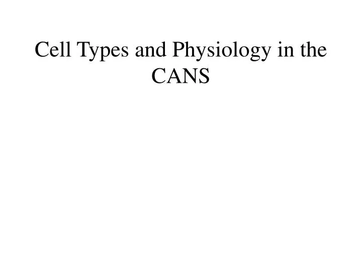
Cell Types and Physiology in the CANS. Major Components of the Central Auditory Nervous System (CANS). VIIIth cranial nerve Cochlear Nucleus Superior Olivary Complex Lateral Lemniscus Inferior Colliculus Medial Geniculate Body Primary Auditory Cortex. Trapezoid body. Brainstem.

E N D
Major Components of the Central Auditory Nervous System (CANS) • VIIIth cranial nerve • Cochlear Nucleus • Superior Olivary Complex • Lateral Lemniscus • Inferior Colliculus • Medial Geniculate Body • Primary Auditory Cortex Trapezoid body Brainstem Mid-brain Thalamus Temporal Lobe
MedGen Body • Inf Coll • Lat Lemn • SOC • Coch Nuc • VIIIth CN
VIIIth Nerve Afferents • Bipolar Neurons • Primary Response Type • Synapse in: • AVCN • PVCN • DCN Sound On Sound Off
Cochlear Nucleus • Wide variety of cell types • AVCN >>to ipsi and contra SOC • PVCN >>to contra Lat. Lemniscus & IC • DCN>>contra to Lat Lemniscus
CN Response Types • Primary-like cell- spherical (bushy) cells of the AVCN • Chopper cell- identification with any particular cell type is not possible because responses are found throughout the cochlear nucleus • Onset- located in octopus cells • Pauser cell/ Build up cell- located in the fusiform layer of the DCN
Pauser cell Onset cell Primary-like cell Buildup cell CN Response Types Chopper cell
DCN Networking • Intranuclear connex • (largely inhibitory) • Enhancing tuning? • Outputs rising in lateral lemniscus • Predominantly contralateral
Superior Olivary Processing Supports Localization • Lateral SO-- Interaural Intensity Differences • Medial SO-- Interaural Time Differences (These are the two primary acoustic cues for localizing sounds)
SOC Physiology • Lateral SOC (EI and predominantly high freq) • Max Response to large SPL diffs between ears • Equal SPLs produce little activation • Larger in animals with smaller heads • Medial SOC (predominantly EE and low freq) • Max Response to specific interaural time diffs • Larger in man than in animals with smaller heads
Lateral Lemniscus • Tract of axons from just above SOC to IC (originating from cell bodies in several different structures) • Has a Nucleus (Nucleus of LL) • Good Temporal Resolution • Involved in Startle Reflex • Connection from CN to Pontine Reticular Formation • PRF >>motor neurons and spinal interneurons
Dorsal (back) Side of Brainstem • Thalamus (medial geniculate) • Inferior Colliculus • 4th Ventricle • Area of Pons
Inferior Colliculus • Cells respond to characteristic: • Interaural delays • Interaural amplitude differences • Amplitude modulations • Frequency modulations • Integration of multi-modal sensory inputs • Adjacent visual nuclei • Proprioception of head and neck • Outputs include oculomotor nuclei • IC stim modifies activity in brain areas involved in attention and learning.
Medial Geniculate Body Thalamus • Last Sensory Relay Station prior to Cortex • Complex of nuclei • MGB is mainly auditory • But has other inputs as well • AND • Some other nuclei respond to aud. stim.
MGB • Three Sub-divisions: • Ventral MGB – True Auditory Relay nucleus • Medial MGB – Auditory with Somatosensory • Dorsal MGB –Somatosensory
Ventral MGB: • Relay cells with overlapping dendritic “nests” • Interneurons inhibit relay cells & other interneurons • Tonotopic Lows-lateral, highs-medial. • Most (90%) Cells are binaural (EE or EI) • 10% Monaural cells: contralateral excitation
Medial MGB: • Cells have CF, • Some show broad tuning • Some have TWO CFs • “Characteristic Intensity” in some cells • EE, EI, IE • All respond for duration of stim • Little Adaptation • But show Habituation • Influenced by somatosensory inputs
Dorsal MGB • Inputs • From IC Both Aud. & Mixed • From Superior Colliculus • Somatosensory Afferents • “Nonspecific” responses
Auditory Radiations Connect • Medial Geniculate Body (in purple) to • Primary Auditory Cortex (in blue)
Primary Auditory Cortex (AI):superior surface of the temporal lobe
6 Cortical Layers • Thalamic inputs >IV • project to pyramidal cells in layer III • Divergence from III • within AI • other cortical areas • contra AI • V and VI >>thalamus &IC
Cortical Neurons • Tonotopic • Lows lateral • Highs medial • Spatiotopic • (best responses from contralateral hemifield) • Strong Habituation/Learning • Sensitive to CHANGES in Frequency and Intensity
Cortical Processing • Pattern Recognition • Duration Discrimination • Localization of Sounds • Selective Attention
Cerebral Dominance • Most right-handed individuals show distribution of language processing in the left hemisphere. (Remember the right ear has the strongest connections to the left hemisphere) • Most people show a right-ear advantage in processing linguistic stimuli