Download
1 / 1

E N D
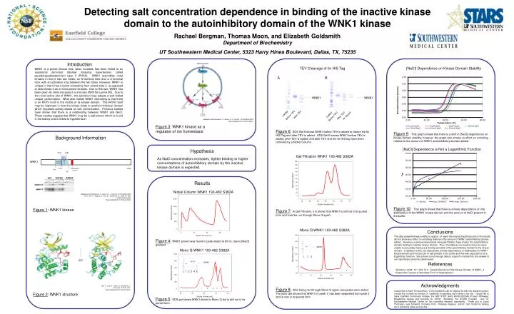
Introduction WNK1 is a protein kinase that, when mutated, has been linked to an autosomal dominant disorder featuring hypertension called psuedohypaldosteronism type II (PHAII). WNK1 resembles most kinases in that it has two lobes, an N-terminal lobe and a C-terminal lobe, with an activation loop between the two lobes. However, WNK1 is unique in that it has a lysine emanating from strand beta 2 as opposed to strand beta 3 as in most protein kinases. Due to this fact, WNK1 has been given its name because it is a kinase (With No Lysine [K]). Due to the novel active site of WNK1, the activation loop adopts a well-folded unique conformation. What also makes WNK1 interesting is that there is an RFXV motif in the middle of its kinase domain. This RFXV motif may be important in how the kinase binds to another inhibitory domain which regulates activity based on salt concentration. Previous studies have shown that there is a relationship between WNK1 and NaCl. These studies suggest that WNK1 may be a salt-sensor which is found in the kidney and is linked to hypertension. Detecting salt concentration dependence in binding of the inactive kinase domain to the autoinhibitory domain of the WNK1 kinase Rachael Bergman, Thomas Moon, and Elizabeth Goldsmith Department of Biochemistry UT Southwestern Medical Center, 5323 Harry Hines Boulevard, Dallas, TX, 75235 S378 S382 P L 1 2 3 4 5 WNK1 Kinase Domain 1 Autoinhibitory Domain 2382 RFXV 194 485 TEV Cleavage of 6x HIS Tag [NaCl] Dependence on Kinase Domain Stability A B WNK1 WNK1 Ladder Ladder After TEV After TEV Ni. Eluant Before TEV Before TEV Adapted from Richardson, C.; Alessi, D.; J. Cell Sci. 121(20)3293-3304 Figure adopted from Thomas Moon Figure 3: WNK1 kinase as a regulator of ion homestasis Figure 6: SDS Gel A shows WNK1 before TEV is added to cleave the 6x HIS Tag and after TEV is added. SDS Gel B shows WNK1 before TEV is added, after TEV is added, and after TEV and the 6x HIS tag have been removed by a Nickel Column. Figure 9: This graph shows that there is a shift in [NaCl] dependence on kinase domain stability, however the graph also shows no effect on unfolding relative to the amount of WNK1 autoinihibitory domain added. Background Information [NaCl] Dependence is Not a Logarithmic Function Hypothesis Gel Filtration WNK1 193-482 S382A As NaCl concentration increases, tighter binding to higher concentrations of autoinhibitory domain by the inactive kinase domain is expected. Results Nickel Column WNK1 193-482 S382A Adapted from Richardson, C.; Alessi, D.; J. Cell Sci. 121(20)3293-3304 Xu, B.; Min, X.; Stippec, S.; Lee, B..; Goldsmith, E.; Cobb, M.; JBC 277(50)48456-48462 Figure adopted from Thomas Moon Figure 10: This graph shows that there is a linear dependence on the stabilization of the WNK1 kinase domain and the amount of NaCl present in the buffer. Figure 1: WNK1 kinase Figure 7: In Gel Filtration, it is shown that WNK1 is still not in its purest form and must be run through Mono Q again. Mono Q WNK1 193-482 S382A Conclusions The data presented was unable to support or reject the original hypothesis since the results did not show any effect on unfolding relative to the amount of WNK1 autoinhibitory domain added . However, previous experiments using gel filtration have shown the autoinhibitory domain binding to inactive kinase domain. Thus, this data is inconclusive since we were unable to accurately measure a binding constant of the autoinhibitory domain to the kinase domain. In addition to this, the data shows a linear dependence on stabilization of WNK1 kinase domain and the amount of salt present in the buffer and this was expected to be a logarithmic function. Since there is not enough data to support or refute this, the answer to our hypothesis cannot be determined. Figure 4: WNK1 protein was found in peak eluted at 60 mL due to [NaCl] gradient. WNK1 Mono Q WNK1 193-482 S382A L 1 2 WNK1 References -Structure, 2004; 12: 1303-1311. Crystal Structure of the Kinase Domain of WNK1, a Kinase that Causes a Hereditary Form of Hypertension 1 2 Acknowledgments Min, X.; Lee, B.; Cobb, M.; Goldsmith, E. Structure 12 1303-1311 Figure adopted from Thomas Moon 1 2 3 4 5 Figure 8: After being ran through Mono Q again, two peaks were eluted. The SDS Gel shows that WNK1 (in peak 1) has been separated from peak 2 and is now in its purest form. I would like to thank Thomas Moon of the Goldsmith Lab for helping me with this research project. I would like to thank my mentor Dr. Goldsmith for allowing me to work in her lab. I would like to thank Eastfield Community College, the NSF STEP Grant #DUE-0525536 (Project Pathways: Broadening Access and Success for STEM Students), the STARS Program, and UT Southwestern Medical Center for this incredible research opportunity. Thank you to Laura Thomason, Judy Schwartz, Professor Post, Professor Hughes, and Dr. Carl Knight for helping me in achieving goals such as this. Figure 2: WNK1 structure Figure 5: SDS gel shows WNK1 eluted in Mono Q but is still not in its purest form.