Download
1 / 21

E N D
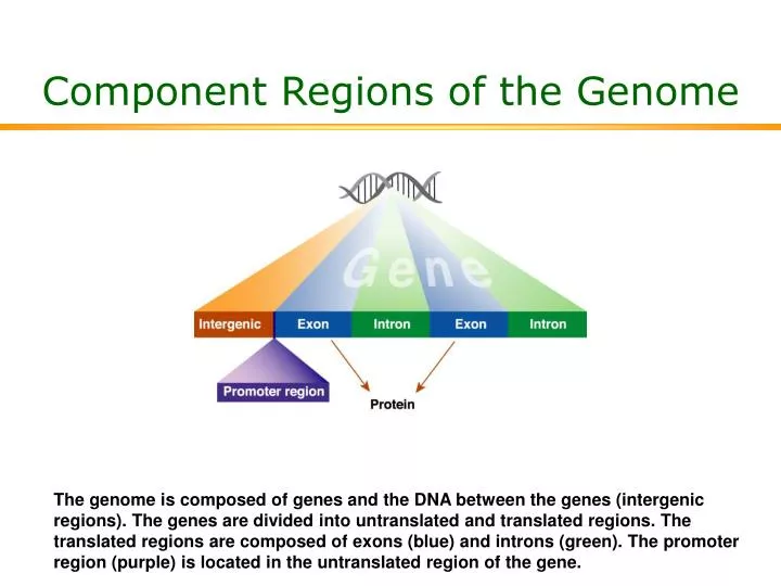
Component Regions of the Genome The genome is composed of genes and the DNA between the genes (intergenic regions). The genes are divided into untranslated and translated regions. The translated regions are composed of exons (blue) and introns (green). The promoter region (purple) is located in the untranslated region of the gene.
Structure of DNA DNA is composed of 2 strands, each composed of a 5-carbon sugar (deoxyribose), a phosphate group (PO4), and a nucleotide base. One strand runs in a 5´ to 3´ direction, while the other runs in the 3´ to 5´ direction. Two of the bases (the purines, shown in blue) are larger than the others (the pyrimidines, shown in green) and are represented by capital letters.
Linkage Disequilibrium Linkage disequilibrium (LD) is a measure of the likelihood of a SNP and an unknown gene to be inherited together. If a SNP is located close to the unknown gene, there is a high LD. If the SNP and the unknown gene are farther apart, there is a greater chance that they will not be inherited together (low LD).
Alternative Splicing The pre-mRNA contains the introns and the exons encoded in the DNA. For the mRNA to produce a functional protein, the introns must be removed. In removing the introns, a variety of potential exon combinations are possible, ie, different combinations of exons may be joined together to generate different forms of the same protein.
Component Regions of RNA The leading and trailing regions, the 5´ and 3´ untranslated regions (UTR), are important for cellular positioning, message stability, and the efficiency with which protein can be made from the mRNA. The open reading frame codes for the protein.
Catabolism of Uracil or Thymine via DPD Uracil is indicated by an H (green) and thymine by CH3 (blue). NADP+ = oxidized form of nicotinamide adenine dinucleotide phosphate; NADPH = reduced form of nicotinamide adenine dinucleotide phosphate.
Pathways for the Anabolic and Catabolic Metabolism of 5-FU dTMP = thymidylate; dUMP = deoxyuridine monophosphate; FBAL = -fluoro-ß-alanine; FdUDP = fluorodeoxyuridine diphosphate; FdUMP = fluorodeoxyuridine monophosphate;FdUTP = fluorodeoxyuridine triphosphate; FUDP = fluorouridine diphosphate; FUdR = fluorodeoxyuridine; FUH2 = dihydrofluorouracil; FUMP = fluorouridine monophosphate; FUPA = -fluoro-ß-ureidopropionate; FUrd = 5-fluorouridine; FUTP = fluorouridine triphosphate; 5-FU = fluorouracil; TS = thymidylate synthetase.
Peripheral Blood Mononuclear (PBM) Cell Method for DPD Activity Assay
5-FU Pharmacokinetics in a DPD-Deficient Patient From Diasio et al. J Clin Invest. 1988;81:47-51, with permission.
Genetic Heritage of a Completely DPD-Deficient Woman A line through a symbol represents deceased members of the family, and the dashed symbols represent members who were alive but not tested for DPD status. The solid symbol represents complete DPD deficiency, the half-filled symbols represent partial deficiency, and the open symbols represent control levels of DPD activity. A double line represents consanguineous marriage. From Diasio et al. J Clin Invest. 1988;81:47-51, with permission.
DPD Activity in a Healthy Population Lu et al. Cancer Res. 1993;53:5433-5438.
Differential Distribution of DPD Activity in Women With Breast Cancer vs Healthy Controls Lu et al. ClinCancer Res. 1998;4:325-329.
Linear Structure of the DPD Protein Schematic of the location of the active site and various domains of the DPD protein. FAD = flavin adenine dinucleotide; Fe-S = iron-sulfur; FMN = flavin mononucleotide; NADP = oxidized form of nicotinamide adenine dinucleotide phosphate; NADPH = reduced form of nicotinamide adenine dinucleotide phosphate.
Stereoview of DPD Protein With Marked Functional Groups FAD = flavin adenine dinucleotide; FeS = iron-sulfur group; FMN = flavin mononucleotide; NADP = nicotinamide adenine dinucleotide phosphate.
Structural Organization of Human DPD Gene (DPYD) Bars represent the exons; intervening spaces represent the introns. Johnson et al. Cancer Res. 1997;57:1660-1663.
Human DPYD Alleles and Location and Relative Frequency of Selected Variants
Variations in the DPD Gene Detected by Allele-Specific PCR One primer (probe) was perfectly complementary to the wild-type (“normal”) sequence at the end of exon 14. The other was perfectly complementary to the sequence for the DPYD*2A variant at the same location. Perfect complementation is necessary for PCR amplification to proceed. Johnson et al. Clin Cancer Res. 1999;5:2006-2011.
Optimization of Running Temperature for DHPLC for DPYD DHPLC = denaturing high-performance liquid chromatography. Ezzeldin et al. Ann Biochem. 2002;306:63-73.
Column Retention Time for Wild-Type and Variant DNA of DPYD by DHPLC Ezzeldin et al. Ann Biochem. 2002;306:63-73.
5-FU Conversion From Prodrug to Active Anabolite or Inactive Metabolite A, 5-FU can enter the metabolic pathway that converts it to fluorodeoxyuridine monophosphate (FdUMP), which can be converted to fluorodeoxyuridine diphosphate (FdUDP), or can inhibit thymidylate synthetase (TS). Inhibiting TS prevents the conversion of deoxyuridine monophosphate (dUMP) to thymidylate (dTMP), blocking the proper formation of DNA. FdUDP can be converted to fluorodeoxyuridine triphosphate (FdUTP), which is added to the growing DNA strands, where it alters the proper functioning of the DNA, leading to cell damage and death. B, 5-FU is converted to fluorouridine diphosphate (FUDP), which can be converted to either fluorouridine triphosphate (FUTP) or FdUDP. C, Most of the administered dose of 5-FU is converted by a series of enzymes (of which DPD is the rate-limiting step) to inactive metabolites dihydrofluorouracil (FUH2), -fluoro-ß-ureidopropionate (FUPA), and -fluoro-ß-alanine (FBAL).