Download
1 / 10
400 likes | 1.87k Vues
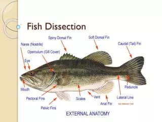
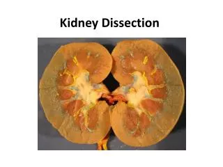
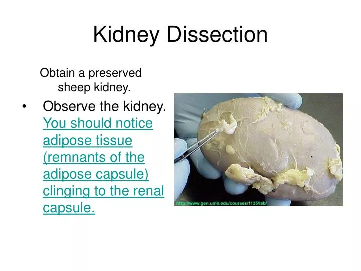
Kidney Dissection. Obtain a preserved sheep kidney. Observe the kidney. You should notice adipose tissue (remnants of the adipose capsule) clinging to the renal capsule. Dissected Kidney 1.Renal Capsule 2. Renal Cortex 3. Renal Medulla 4.Renal Pyramid 5.Renal Pelvis 6.Renal Column

E N D
Kidney Dissection Obtain a preserved sheep kidney. • Observe the kidney. You should notice adipose tissue (remnants of the adipose capsule) clinging to the renal capsule.
Dissected Kidney • 1.Renal Capsule • 2. Renal Cortex • 3. Renal Medulla • 4.Renal Pyramid • 5.Renal Pelvis • 6.Renal Column • 7.Renal Calyx • 8.Ureter
You should also notice a "pinched-in" area where the renal blood vessels and ureter are attached to the kidney. This is the renal hilus.
Remove the renal capsule by carefully cutting through it and grabbing the cut capsule with forceps.
Once the renal capsule is removed, you will be looking at the renal cortex • In order to more accurately identify each, separate the renal blood vessels from one another and from the ureter.
Generally, the tube with the most adipose around it is the ureter. Notice the histological differences (and similarities) between the renal arteries, renal veins, and ureter.
Carefully make a frontal (coronal) section through the kidney.
Identify as many parts of the internal structure of the kidney as possible (renal cortex, renal medulla, renal pelvis, etc.). Recall the reading and discussion of kidney function and think about this as you observe kidney anatomy.