Download
1 / 1
10 likes | 352 Vues
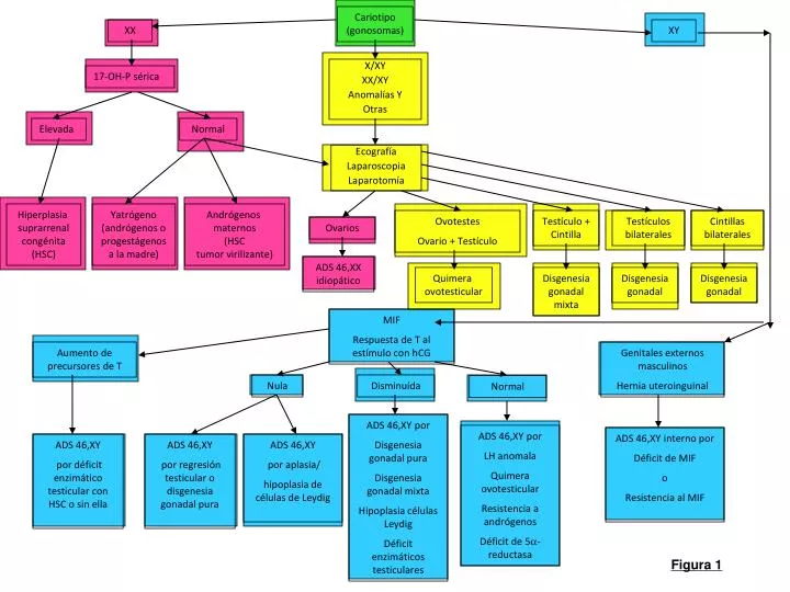
Cariotipo (gonosomas). XX. XY. X/XY XX/XY Anomalías Y Otras. 17-OH-P sérica. Elevada. Normal. Ecografía Laparoscopia Laparotomía. Hiperplasia suprarrenal congénita (HSC). Yatrógeno (andrógenos o progestágenos a la madre). Andrógenos maternos (HSC tumor virilizante).

E N D
Cariotipo (gonosomas) XX XY X/XY XX/XY Anomalías Y Otras 17-OH-P sérica Elevada Normal Ecografía Laparoscopia Laparotomía Hiperplasia suprarrenal congénita (HSC) Yatrógeno (andrógenos o progestágenos a la madre) Andrógenos maternos (HSC tumor virilizante) Ovotestes Ovario + Testículo Testículo + Cintilla Testículos bilaterales Cintillas bilaterales Ovarios ADS 46,XX idiopático Quimera ovotesticular Disgenesia gonadal mixta Disgenesia gonadal Disgenesia gonadal MIF Respuesta de T al estímulo con hCG Aumento de precursores de T Genitales externos masculinos Hernia uteroinguinal Nula Disminuída Normal ADS 46,XY por Disgenesia gonadal pura Disgenesia gonadal mixta Hipoplasia células Leydig Déficit enzimáticos testiculares ADS 46,XY por LH anomala Quimera ovotesticular Resistencia a andrógenos Déficit de 5a-reductasa ADS 46,XY interno por Déficit de MIF o Resistencia al MIF ADS 46,XY por déficit enzimático testicular con HSC o sin ella ADS 46,XY por regresión testicular o disgenesia gonadal pura ADS 46,XY por aplasia/ hipoplasia de células de Leydig Figura 1