Download
1 / 11
110 likes | 288 Vues
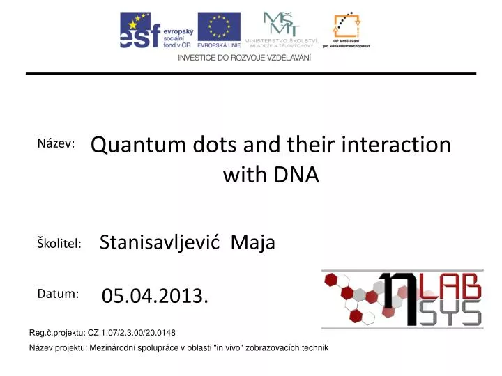
Název: Školitel: Datum:. Quantum dots and their interaction with DNA. Stanisavljević Maja. 05.04.2013. Reg.č.projektu : CZ.1.07/2.3.00/20.0148 Název projektu: Mezinárodní spolupráce v oblasti "in vivo " zobrazovacích technik. Quantum dots – history of synthesis

E N D
Název: Školitel: Datum: Quantum dots and their interaction with DNA StanisavljevićMaja 05.04.2013. Reg.č.projektu: CZ.1.07/2.3.00/20.0148 Název projektu: Mezinárodní spolupráce v oblasti "in vivo" zobrazovacích technik
Quantum dots – history of synthesis • 1980 – Ekimov, Efros – first description • of quantum dots • 1993 - Murray, Norris, Bawendi • CdE(E=S,Se,Te) and other improvments • like ZnS shell • Semiconductor nanocrystals synthetized • from II and VI or III and V elements of PSE. • today-enviromental-friendly synthesis ZnS CdTe • Characteristics • small size 1-10nm • absorbtion of light in wide range • emmision range very narrow • photostability and resistance to • chemical degradation • poisonous core and hydrophobic • surface modification • bioconjugation
DNA • 1869 – isolated by Swiss scientist • Friedrich Miescher • 1953 – described DNA structure • Watson and Crick • 1962 – they recieved Nobel prize • for solving nature’s biggest secret • DNA helix-two complementary and antiparallel polynucleotide strands QD • Experiment hypothesis: • Major groove – 2.2 nm • QD-GSH – 2 nm • DNA invisible for CE-LIF • Mix DNA and observe
Measurements conditions • CE-LIF • Capillary lenght: ltot=57cm,leff=50cm, ID=75µm • BGE: 20mM borate pH 9.2 • Separation: 20kV, positive polarity • Injection: 3.4kPa for 20sec • Excitation wavelenght: 488nm • Emmision wavelenght: 520 nm • Sample: QD-GSH and DNA from chicken in conc. of 1 mg/mL
QD-GSH λex = 380 nm λem = 524 nm If (a.u.) CE-LIF QD-GSH Migration time (min) If (a.u) heated QDs 10 min at 50oC Intensity % Migration time (min) Size (nm)
QD-GSH + DNA – interaction through time IF (a.u.) QD Migrationtime (min) QD
QD-GSH with different concentration of DNA DNA 1 – 0.25 mg/mL DNA 2 – 0.50 mg/mL DNA 3 – 1.00 mg/mL QD-GSH + DNA 3 QD-GSH + DNA 2 IF (a.u.) QD-GSH + DNA 1 QD-GSH Height of peak Time of migration (min) Concentraton of DNA (mg/mL)
QD-GSH - dsDNAvsssDNA ssDNA IF (a.u.) No complex dsDNA QD-GSH - hybridization of DNA QDs + ssDNA QDs + dsDNA QDs Migrationtime (min) IF (a.u.) Hybridization of DNK: 99oC, 15 min Migrationtime (min)
Conclusions • QDs and dsDNA interacts creating complex – in shape of peak • Interaction peak grows through time and increasing DNA concentration • Complex is not created with ssDNA or hybridized dsDNA– possible confirmation of QDs being caught in major groove • Some QDs with different capping agents
Thanks to : • Departament of chemistry and biochemistry • Jana Chomoucka for QD preparing
THANK YOU ATTENTION! FOR