Download
1 / 1
10 likes | 212 Vues
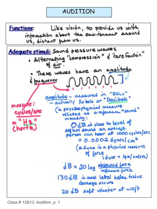
Audition. Transduction of Mechanical Displacement. Physiology of the Cochlea. Generation of Action Potential (AP). Auditory Pathway. Cochlea. Basic Auditory Pathway. Higher Brain Activities Involved in Auditory Perception. Localization of Sound. Coding of Intensity and Frequency.

E N D
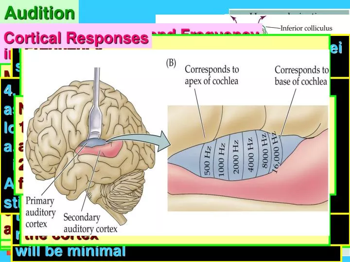
Audition Transduction of Mechanical Displacement Physiology of the Cochlea Generation of Action Potential (AP) Auditory Pathway Cochlea Basic Auditory Pathway Higher Brain Activities Involved in Auditory Perception Localization of Sound Coding of Intensity and Frequency Background & Structure of Auditory System Cortical Responses 1. Dorsal and posteroventral cochlear nuclei send projections to the inferior colliculus a. Pathways respond to sound arriving at one ear only 2. Auditory maps a. Auditory space is not mapped at the cortical level. Tonotopic maps are created at the levels of the inferior colliculus 3. Highly processed auditory information is then relayed to the medial geniculate nucleus of the thalamus Frequency 1. Consequence of the mechanics of the basilar membrane 2. Different portions of the basilar membrane are maximally deformed by sound of different frequencies 3. Hair cells that are selectively activated project to cochlear nuclei (brain stem) with tonographic specificity a. Specificity is conserved all the way to the cortex Transduction 1. Bending of these cilia is the critical event in the transduction of sound into neural signal 2. Hairs extend above the reticular membrane and come in contact with tectorial membrane 3. When the basilar membrane moves in response to the motion of the stapes a. Whole complex moves as a unit either towards or away from the tectorial membrane b. Lateral motion of the reticular membrane bends the stereocilia Inner ear Middle ear Localization of sounds above 3 kHz Circuit 1. Medial superior olive (MSO) has cells that receive coincident innervation from the right and left anteroventral cochlear nucleus a. Cells within the MSO are organized such that the distance from the respective cochlear nuclei varies systematically i. Length of the axonal connections determine which MSO cell receives coincident activation by action potential 6. Depending on the direction that hairs bend, the channel will either be opened or closed a. Opening the channel allows K+ to enter and depolarize the hair cell b. Closing the channel stops the flow of K+ 7. In response to be depolarization resulting from influx of K+ a. Ca++ channel is activated b. Influx of Ca++ causes the release of synaptic vesicles from the end of the hair cell Note: Subjects need to popped their ears when they go up in an airplane. On the ground their middle ear is as the same pressure as the outside environment. As they ascend, air pressure is lower a high altitudes. The tympanic membrane will bulge out because the pressure in the middle ear is greater than the outside environment. When they yawn or swallow, the valve in the tube is opened and the pressure is relieved. 4. Mechanism: Anteroventral cochlear nuclear firing rate is greater for sound with higher intensities i. Sound arising directly lateral to the listener, LSO firing will be highest on that side ii. Excitation from the ipsilateral anteroventral cochlear nucleus will be maximal iii. Inhibition from the contralateral MNTB will be minimal c. Bones in the middle ear amplify the pressure. Pivot points that act as fulcrums. Malleus is displaced in response to the movement of the tympanic membrane--bottom moves towards the inner ear and the top moves towards the outer ear. This pulls the top of the incus towards the outer ear and pushes the bottom towards the inner ear. Stapes is consequently pushed forward against the oval window which is compressed inward Organ of Corti 1. Structure a. Outer hair cells b. Inner hair cells c. Tectorial membrane d. Reticular membrane e. Basilar membrane f. Stereocilia g. Spiral ganglion AP occurs at the level of output ganglion 1. Multiple outer hair cells make synaptic contact with a single ganglion cell. 2. Ganglion make synaptic contact with a single inner hair cell (although many ganglion cells can contact the same inner hair cell) 3. 75% of all hair cells are outer hair cells a. Outer alter the stiffness of tectorial membrane 4. Only 5% of the fibers in the auditory nerve are from outer hair cells Connections to the brain stem 1. Spiral ganglion sends projection to the cochlear nucleus a. There are two cochlea, each projecting to its cochlear nucleus b. Within the cochlear nucleus this process branches i. Dorsal cochlear nucleus ii. Posterior ventral cochlear nucleus iii. Anterior ventral nucleus • Processes • Sound waves move the • tympanic membrane. • 2. Tympanic membrane moves • the ossicles. • 3. Ossicles move the membrane • at the oval window. • 4. Motion at the oval window • moves the fluid in the cochlea. 4. Depending on the direction that the hairs bend, the inside of the hair cells will either: a. Depolarize b. Hyperpolarize 5. Changes in cell potential result from the opening of K+ channels on tips of stereocilia a. Channels are mechanically gated b. Flaps that are connected to neighboring cilia by a special protein molecule • Outer ear • 1. Pinna • Funnel shaped outer ear made • of skin and cartilage • 2. Auditory canal • Channel leading from the pinna • to the tympanic membrane Monoaural systems 2. Ossicles: Series of bones in a small air filled chamber. Transfer the movement of the tympanic membrane into the movement of a second membrane covering a hole in the bone of the skull (oval window). The bones of middle ear are malleus (hammer), incus (anvil) and stapes (stirrup) c. Dorsal and posterior ventral cochlear nuclei send efferent projections to the contralateral inferior colliculus i. Via the nucleus of the lateral leminiscus d. Anterior ventral cochlear nucleus is a critical component of a brainstem neural circuit that permits the detection of interaural time differences 3. Basilar membrane a. Separates scala media and scala tympani b. Properties are very important for audition 4. Fluid is continuous between scala vestibuli and scala tympani a. Physical connection is known as the helicotrema • Process • 1. Mechanical forcepusheson the oval window • 2. Fluid within the cochlea is incompressible • 3. Fluid pushes forward • Conserves the wave properties of thesound • (i.e. the movement of the fluid has frequency • and amplitude) • b. Causes the round window to bulge out • Sound • Audible variations in air • pressure (compressions) • 2. Molecules are displaced • forward leaving a corresponding • area of lower pressure 3. Circuit: Anteroventral cochlear nucleus projects directly to the ipsalateral lateral superior olive (LSO) i. Indirectly to the contralateral lateral superior olive via an inhibitory neuron originating in the medial nucleus of the trapezoid body (MNTB) c. High frequency sounds have higher energy and can displace the stiffer part of the basilar membrane (near the base) d. Lower frequency sounds have lower energy and displace the apex end e. Base responds to high frequency and the apex responds to low frequency Sequence overview: 1. Physical displacement of the basilar membrane bends the stereocilia 2.Bending of cilia opens or closes K+ channel 3. When K+ enters, the hair cell depolarizes 4. Depolarization activates a Ca++ channel 5. Ca++ influx causes NT release 4. From the thalamus, this information ascends to the primary auditory cortex (A1) located in the temporal lobe a. A1 has a topographical map of the cochlea i. Specific regions (isofrequency bands) of A1 are activated in response to acoustical stimulation of the basilar membrane 5. Structural properties of the basilar membrane determine the way it responds to sound a. Membrane is wider at apex than base (5:1) b. Stiffness of the membrane decreases from base to apex (like a diving board) Anatomy 1. Cross section 2. Chambers of the cochlea a. Scala vestibuli b. Scala tympani c. Scala media Audition 1. Sense of hearing 2. Mechanisms within the ear and brain that translate sound in our environment into meaningful neural signals • Converts the physical movement of the oval window into neural signal 2. Takes place in the cochlea 3. Elements a. Cochlea b. Vestibular apparatus i. Not part of the auditory system ii. Involved in balance Intensity 1. Firing rate of individual hair cells 2. Activation of multiple hair cells. 3. Wave of higher amplitude has more width and activates more hair cells in a given area Mechanisms for detecting interaural time differences 1. Two ears separated by about 20 cm i. Diameter of your head 2. Detect differences as small as 10 msec 3. Functional considerations: a. Cochlea is filled with an incompressible fluid b. More force is required to displace fluid than air d. Oval window is smaller and the same pressure across a smaller area results in a greater force (like a spiked high heel) 4. Eustachian tube: Tube that connects the air-filled middle ear to the mouth. It Contains a valve • 3. Sound waves vary in two ways: • Amplitude--intensity; peak to trough; • perceived as differences in loudness • b. Frequency: Number of compressions per • second; pitch; unit: hertz (1 cycle/second) Three divisions of the ear 1. Outer 2. Middle 3. Inner 1. Sound does not bend around the head 2. Directed to one side or the other and an intensity difference results Neuronal organization 1. Isofrequency bands a. Temporal lobe 2. Neurons within these bands respond to fairly similar characteristic frequencies Background Cochlea transduces the mechanical displacement of the oval window into a neural signal 6. Basilar membrane establishes a place code in which different locations are maximally deformed in response to different frequency sounds 5. Movement of the fluid in the cochlea causes a response in sensory neurons. 6. Signal is transferred and processed by a series of nuclei in the brain stem. • 1. Tympanic membrane (eardrum) • Moves in response to variations • in air pressure 4. Structures within the cochlea are not rigid. Basilar membrane is flexible and bends in response to sound 7. Information is sent to a relay in the thalamus(medial geniculate nucleus-MGN) 8. MGN projects to the primary auditory cortex in the temporal lobe. 3. Organ of corti a. Contains auditory receptor cells b. Located in the scala media Auditory receptors 1. Hair cells a. Stereocilia Limitations 1. System works well for sounds that have frequencies below 3 kHz Home Exit BASIM ZWAIN LECTURE NOTES 4. From the thalamus, this information ascends to the primary auditory cortex (A1) located in the temporal lobe