Download
1 / 1
30 likes | 181 Vues
Surface Reconstruction of Blood Vessels from 3D Fluorescence Microscopy Images. Arunachalam Narayanaswamy * , Badrinath Roysam*, Barbera Cutler ¤ , Chris Bjornsson § and William Shain ¶

E N D
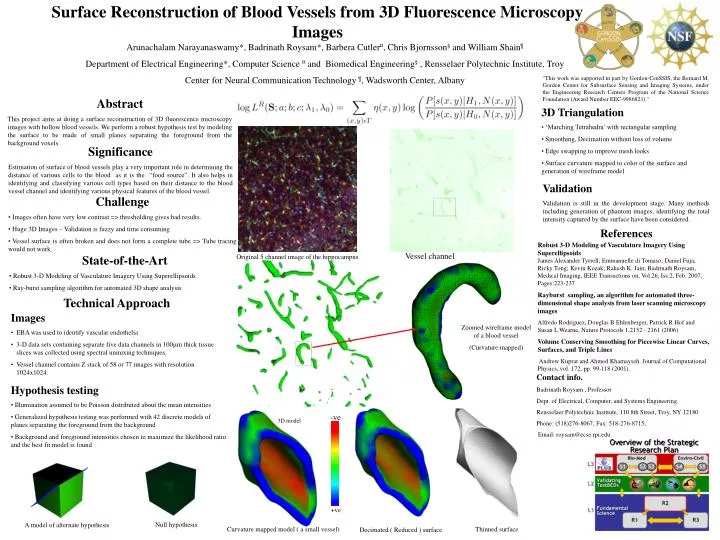
Surface Reconstruction of Blood Vessels from 3D Fluorescence Microscopy Images Arunachalam Narayanaswamy*, Badrinath Roysam*, Barbera Cutler¤, Chris Bjornsson§ and William Shain¶ Department of Electrical Engineering*, Computer Science ¤ and Biomedical Engineering§ , Rensselaer Polytechnic Institute, Troy Center for Neural Communication Technology ¶, Wadsworth Center, Albany "This work was supported in part by Gordon-CenSSIS, the Bernard M. Gordon Center for Subsurface Sensing and Imaging Systems, under the Engineering Research Centers Program of the National Science Foundation (Award Number EEC-9986821)." Abstract This project aims at doing a surface reconstruction of 3D fluorescence microscopy images with hollow blood vessels. We perform a robust hypothesis test by modeling the surface to be made of small planes separating the foreground from the background voxels. • 3D Triangulation • ‘Marching Tetrahedra’ with rectangular sampling • Smoothing, Decimation without loss of volume • Edge swapping to improve mesh looks • Surface curvature mapped to color of the surface and generation of wireframe model Significance Estimation of surface of blood vessels play a very important role in determining the distance of various cells to the blood as it is the “food source”. It also helps in identifying and classifying various cell types based on their distance to the blood vessel channel and identifying various physical features of the blood vessel. Validation Validation is still in the development stage. Many methods including generation of phantom images, identifying the total intensity captured by the surface have been considered. • Challenge • Images often have very low contrast => thresholding gives bad results. • Huge 3D Images – Validation is fuzzy and time consuming • Vessel surface is often broken and does not form a complete tube => Tube tracing would not work. References Robust 3-D Modeling of Vasculature Imagery Using SuperellipsoidsJames Alexander Tyrrell; Emmanuelle di Tomaso; Daniel Fuja; Ricky Tong; Kevin Kozak; Rakesh K. Jain; Badrinath Roysam, Medical Imaging, IEEE Transactions on, Vol.26, Iss.2, Feb. 2007, Pages:223-237 Rayburst sampling, an algorithm for automated three-dimensional shape analysis from laser scanning microscopy images Alfredo Rodriguez, Douglas B Ehlenberger, Patrick R Hof and Susan L Wearne, Nature Protocols 1,2152 - 2161 (2006) Volume Conserving Smoothing for Piecewise Linear Curves, Surfaces, and Triple Lines Andrew Kuprat and Ahmed Khamayseh. Journal of Computational Physics, vol. 172, pp. 99-118 (2001). Vessel channel • State-of-the-Art • Robust 3-D Modeling of Vasculature Imagery Using Superellipsoids • Ray-burst sampling algorithm for automated 3D shape analysis Original 5 channel image of the hippocampus Technical Approach • Images • EBA was used to identify vascular endothelia • 3-D data sets containing separate five data channels in 100m thick tissue slices was collected using spectral unmixing techniques. • Vessel channel contains Z stack of 58 or 77 images with resolution 1024x1024. Zoomed wireframe model of a blood vessel (Curvature mapped) Contact info. Badrinath Roysam , Professor Dept. of Electrical, Computer, and Systems Engineering Rensselaer Polytechnic Institute, 110 8th Street, Troy, NY 12180 Phone: (518)276-8067; Fax: 518-276-8715; Email: roysam@ecse.rpi.edu • Hypothesis testing • Illumination assumed to be Poisson distributed about the mean intensities • Generalized hypothesis testing was performed with 42 discrete models of planes separating the foreground from the background • Background and foreground intensities chosen to maximize the likelihood ratio and the best fit model is found -ve 3D model +ve Null hypothesis A model of alternate hypothesis Curvature mapped model ( a small vessel) Thinned surface Decimated ( Reduced ) surface