Download
1 / 68
680 likes | 782 Vues
The Skeletal System. Chapter 5. 1. Objectives:. •List and describe the major functions of the skeletal system. •Describe the types of various bone tissues, their locations, and functions. •List, describe and give specific examples of the types of bones.

E N D
The Skeletal System Chapter 5 1
Objectives: • •List and describe the major functions of the skeletal • system. • •Describe the types of various bone tissues, their • locations, and functions. • •List, describe and give specific examples of the • types of bones. • •List and describe the two major divisions of the • skeletal system and the bones which compose each. • •Describe the process of bone formation. • •List and describe the various types of joints. • •List and describe various disorders and diseases of • the skeletal system. 2
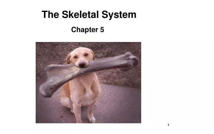
The Skeletal System What would you look like if you did not have any bones? Take a minute a make a sketch of what you think you might look like. Could you stand up? Could you walk? Without bones you would be just a puddle of skin and organs on the floor. Click on the trunk's lid and then use the red knob to pull the object from the trunk.
The human skeleton contains 206 bones!
Functions of the Skeletal System • •Support of the body • •Protection of soft organs • •Movement due to attached skeletal • muscles • •Storage of minerals and fats • •Blood cell formation (hematopoiesis) 3
Types of Bone Tissue ·The skeleton has 206 bones ·Two basic types of bone tissue ·Compact bone ·Homogeneous ·Located on the outside surface ·Spongy bone ·Small needle-like ·pieces of bone (trabeculae) ·Many open spaces Where marrow is located Diagram on p. 118 4
Compact Bone Spongy Bone - hard bone - surrounds spongy bone - made of osteon systems that contain blood vessels - made of osteocytes (living bone cells) - less dense bone - contains many holes and spaces spongy compact
Classification of Bones by Shape •Long bones –Typically longer than wide –Have a shaft (long, central cylinder) with heads at both ends –Contain mostly compact bone •Examples: Femur, humerus •Short bones –Generally cube-shape –Contain mostly spongy bone •Examples: Carpals, tarsals 5
Classification of Bones by Shape •Flat bones –Thin and flattened –Usually curved –Thin layers of compact bone around a layer of spongy bone •Examples: Skull, ribs, sternum •Irregular bones –Irregular shape –Usually with projections (processes) or openings (foramen) –Do not fit into other bone classification categories •Example: Vertebrae and pelvic bones 6
Classification of Bones by Shape Diagram on p. 117 7
Color in the types of bones based on the correct classifications.
Microscopic Bone Anatomy Diagram on p. 120 8
Microscopic Bone Anatomy •Osteon (Haversian System) –A unit of bone •Central (Haversian) canal –Opening in the center of an osteon –Carries blood vessels and nerves •Perforating (Volkman’s) canal –Canal perpendicular to the central canal –Carries blood vessels and nerves Diagram on p. 120 9
Microscopic Bone Anatomy •Lacunae –Cavities containing bone cells (osteocytes) –Arranged in concentric rings •Lamellae –Rings around the central canal –Sites of lacunae •Canaliculi –Tiny canals –Radiate from the central canal to lacunae –Form a transport system between cells Diagram on p. 120 10
Bone Cell Types •Osteocytes –Mature bone cells surrounded by matrix •Osteoblasts –Bone-forming cells produce new matrix •Osteoclasts –Bone-destroying cells –Break down bone matrix for remodeling and release of calcium •Bone remodeling is a process by both osteoblasts and osteoclasts. –During your life span the your skeleton is broken down and rebuilt completely on the average of seven times! 11
Canaliculi Lamella Sharpey's fibers Lacunae Blood vessel Haversian/central canal Osteocytes Periosteum Osteon Compact bone Lamella Volkmann's canal Spongy bone
Review! Bone Classifications Long Bones Flat Bones Irregular Bones Short Bones
Gross Anatomy of a Long Bone Diagram on p. 118 •Diaphysis –Shaft –Composed of compact bone •Epiphysis –Ends of the bone –Composed mostly of spongy bone –Contains red marrow •Medullary cavity –Cavity of the shaft –Contains yellow marrow (mostly fat) in adults •Articular cartilage –Covers the external surface of the epiphyses –Made of hyaline cartilage –Decreases friction at joint surfaces 13
Gross Anatomy of a Long Bone Diagram on p. 118 •Periosteum –Outside covering of the diaphysis –Fibrous connective tissue membrane •Sharpey’s fibers –Secure periosteum to underlying bone •Endosteum - Inner lining of the medullary cavity •Arteries, veins, nerves –Found in the Haversian and Volkman canal systems within the compact bone. 14
Bone Development and Growth •During development, cartilage is replaced by bone (ossification) •Cartilage remains in isolated areas –Bridge of the nose –Parts of ribs –Joints •Bones are remodeled and lengthened until growth stops –Bones change shape somewhat –Bones grow in width Diagram on p. 121 15
Bone Development and Growth •Epiphyseal plates allow for growth of long bone during childhood –New cartilage is continuously formed –Older cartilage becomes ossified •Cartilage is broken down •Bone replaces cartilage 16
Periosteum Compact bone Cartilage Epiphyseal line Medullary Cavity Cartilage Bone marrow Diaphysis Compact bone Compact bone Epiphysis Periosteum Spongy bone Epiphysis Blood vessels Sharpey's fibers Spongy bone
Divisions of the Skeleton •The Skeleton is divided into two major regions: a. Axial Skeleton b. Appendicular Skeleton •Axial Skeleton - longitudinal part of the body –Divided into three parts •Skull •Vertebral column •Bony thorax 19
Divisions of the Skeleton •Appendicular skeleton - appendages and joints –Limbs (appendages) –Pectoral girdle (shoulder, scapula, and clavicle) –Pelvic girdle (pelvis) 20
Divisions of the Skeleton Diagram p. 125 21
Label Mr. Bones! Tibia Cranium Radius Sternum Sacrum Metacarpals Patella Fibula Humerus Ulna Clavicle Mandible Scapula Ribs Pelvic Metatarsals Vertebra Femur
patella humerus femur scapula radius vertebra metatarsals skull clavicle ulna tibia fibula pelvis metacarpals costals
Boneyard Bill's Body Parts Can you drag all the bones to their correct locations?
BONES, BONES, BONES As you find the bones, identify them on Steve! S T E R N U M Y E S J A W B O N E C L A V I C L E G O L S C H O O L R W P R S C A U P U L A B O A T E A B E A T H T S E D P R U X L K P N M A M Z C A I P A T E L L A S H I C L S S A F D O G I A I M U D A U A A B U E T E N E B S E I S R R M T N T R L S A M E I T D A H A B A A G S E A N O T U A A M C L G E N L E R M S E A L A R I B S D O T Y O S E U T A N T N E N I P S N R K B N L H V A L O S T B A N O F E M O S T B E E J S I V L E P F A V How fast can your class find the words? PELVIS TIBIA CLAVICLE CRANIUM PHALANGES ULNA FEMUR RADIUS VERTEBRA FIBULA RIBS SCAPULA HUMERUS JAWBONE STERNUM SPINE PATELLA
Axial Skeleton: The Skull •Two sets of bones –Cranium –Facial bones •Bones are joined by sutures (fixed, non-movable joints) •Only the mandible is attached by a freely movable joint 23
Axial Skeleton: The Skull 24 Diagram p. 126
Diagram p. 129 Axial Skeleton: The Skull 25
Axial Skeleton: The Skull Diagram p. 127 26
Axial Skeleton: The Skull •Paranasal sinuses: Hollow portions of bones surrounding the nasal cavity –Functions •Lighten the skull •Give resonance and amplification to voice Diagram p. 128 27
Auditory meatus Mandibular ramus Mental foramen Frontal bone Nasal bone Ethmoid bone Lacrimal bone Maxilla Suture Temporal bone Mastoid process Occipital bone Sphenoid bone Suture Mandible Suture Zygomatic bone Styloid process Parietal bone
Practice Makes Perfect! Parietal bone Nasal bone Zygomatic bone Temporal bone Optic canal Inferior nasal concha Middle nasal concha Suture Ethmoid bone Lacrimal bone Mandible Vomer Alveolar margins Sphenoid bone Maxilla Frontal bone
Axial Skeleton: Hyoid Bone Diagram p. 130 • ·The only bone • that does not • articulate with • another bone • ·Serves as a • moveable base for • the tongue Figure 5.12 30
Axial Skeleton: Vertebral Column Diagram p. 131 • ·Vertebrae are • separated by • intervertebral discs • composed of • fibrocartilage • ·The spine has a • normal curvature • ·Each vertebrae is • given a name • according to its • location 31
Axial Skeleton: Vertebral Column 32 Diagram p. 133
Thoracic vertebrae Cervical vertebrae C1Atlas Sacrum Coccyx Lumbar vertebrae C2 Axis
Appendicular Skeleton: Pectoral Girdle •Composed of two bones –Clavicle – collarbone –Scapula – shoulder blade •These bones allow the upper limb to have exceptionally free movement 36
Appendicular Skeleton: Pectoral Girdle (Shoulder) 37 Diagram p. 139
Appendicular Skeleton: Brachium Region ·The arm is formed by a single bone ·Humerus Diagram p. 140 38
Appendicular Skeleton: Antebrachium Region •The forearm has two bones •Ulna (side adjacent to little finger) •Radius (side adjacent to thumb) 39 Diagram p. 140
Appendicular Skeleton: Carpus, Manus, and Digits Region ·The hand ·Carpals – wrist ·Metacarpals – palm ·Phalanges – fingers 40 Diagram p. 141
Appendicular Skeleton: Pelvic Girdle •Hip bones •Composed of three pair of fused bones –Ilium –Ischium –Pubic bone •The total weight of the upper body rests on the pelvis •Protects several organs –Reproductive organs –Urinary bladder –Part of the large intestine 41
Appendicular Skeleton: Pelvic Girdle The sacrum and coccyx are part of the axial skeleton not appendicular skeleton. 42 Diagram p. 142
Appendicular Skeleton: Pelvic Girdle Coxal Bone Structure 43 Diagram p. 142
Appendicular Skeleton: Pelvic Girdle Male and Female Pelvis Comparison Diagram p. 142 44