Download
1 / 80
820 likes | 1.59k Vues
Membrane Transport and Membrane Potentials. Dr. Michael P. Gillespie. Action Potential. Have the ability to produce action potentials or impulses (electrical excitability) in response to a stimulus.

E N D
Membrane Transport and Membrane Potentials Dr. Michael P. Gillespie
Action Potential • Have the ability to produce action potentials or impulses (electrical excitability) in response to a stimulus. • An action potential is an electrical signal that propagates from one point to the next along the plasma membrane of a neuron. • A stimulus is any change in the environment that is strong enough to initiate an action potential.
Parts of a Neuron • Cell Body • Dendrites • Axon
Parts of a Neuron (Cell Body) • Cell body (perikaryon or soma). • Contains the nucleus surrounded by cytoplasm which contains the organelles. • Clusters of rough ER called Nissl bodies (produce proteins to grow and repair damaged nerves)
Parts of a Neuron (Nerve Fiber) • Nerve fiber – any neuronal process that emerges from the cell body of a neuron. • Dendrites • Axon
Parts of a Neuron (Dendrites) • Dendrites (= little trees). • The receiving (input) portion of a neuron. • Short, tapering, and highly branched.
Parts of a Neuron (Axon) • Axon (= axis). • Each nerve contains a single axon. • The axon propagates nerve impulses toward another neuron, muscle fiber, or gland cell. • Long, thin, cylindrical projection that often joins the cell body at a cone-shaped elevation called the axon hillock (= small hill). • The part of the axon closest to the hillock is the initial segment. • The junction between the axon hillock and the initial segment is the trigger zone (nerve impulses arise here). • The cytoplasm of the axon is the axoplasm and is surrounded by a plasma membrane known as the axolemma (lemma = sheath).
Synapse • The synapse is the site of communication between two neurons or between a neuron and an effector cell. • Synaptic end bulbs and varicosities contain synaptic vesicles that store a chemical neurotransmitter.
Myelination • The myelin sheath is a lipid and protein covering. It is produced by the neuroglia. • The sheath electrically insulates the axon of a neuron. • The sheath increases the speed of nerve impulse conduction. • The amount of myelin increases from birth on. • Axons without a covering are unmyelinated. Axons with a covering are myelinated.
Myelination Continued… • Two types of neuroglial cells produce myelination. • Schwann cells – located in the PNS. • Oligodendrocytes – located in the CNS.
Neurolemma (Sheath of Schwann) • The neurolemma (sheath of Schwann) is the outer nucleated cytoplasmic layer of the Schwann cell. • It encloses the myelin sheath. • It is only found around the axons of the PNS. • If the axon is injured, the neurolemma forms a regeneration tube that guides and stimulates re-growth of the axon.
Nodes of Ranvier • The nodes of Ranvier are gaps in the myelin sheath at intervals along the axon. • Each Schwann cell wraps one axon segment between two nodes. • The electrical impulse jumps from node to node to speed up the propagation • Nodes of Ranvier are present in the CNS, but fewer in number.
Demyelination • Demyelination is the loss or destruction of the myelin sheaths around axons. • It occurs as the result of disorders such as multiple sclerosis or Tay-Sachs disease. • Radiation and chemotherapy can also damage the myelin sheath. • Demyelination can deteriorate the affected nerves.
Electrical Signals in Neurons • Neurons are electrically excitable and communicate with one another using 2 types of electrical signals. • Graded potentials (short distance communication). • Action potentials ((long distance communication). • The plasma membrane exhibits a membrane potential. The membrane potential is an electrical voltage difference across the membrane.
Electrical Signals in Neurons • The voltage is termed the resting membrane potential. • The flow of charged particles across the membrane is called current. • In living cells, the flow of ions constitutes the electrical current.
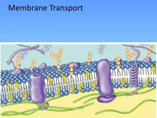
Ion Channels • The plasma membrane contains many different kinds of ion channels. • The lipid bilayer of the plasma membrane is a good electrical insulator. • The main paths for flow of current across the membrane are ion channels.
Ion Channels • When ion channels are open, they allow specific ions to move across the plasma membrane down their electrochemical gradient. • Ions move from greater areas of concentration to lesser areas of concentration. • Positively charged cations move towards a negatively charged area and negatively charged anions move towards a positively charged area. • As they move, they change the membrane potential.
Ion Channel “Gates” • Ion channels open and close due to the presence of “gates”. • The gate is part of a channel protein that can seal the channel pore shut or move aside to open the pore.
Types of Ion Channels • Leakage channels • Ligand-gated channel • Mechanically gated channel • Voltage gated channel
Leakage Channels • Leakage channels – gates randomly alternate between open and closed positions. • More potassium ion (K+) leakage channels than sodium (Na+) leakage channels. • The potassium ion leakage channels are leakier than the sodium ion leakage channels.
Ligand-gated Channel • Ligand-gated channels – open and close in response to a specific chemical stimulus. • Neurotransmitters, hormones, and certain ions can act as the chemical stimulus that opens or closes these channels.
Mechanically Gated Channel • Mechanically gated channels – opens or closes in response to mechanical stimulation. • Vibration, touch, pressure, or tissue stretching can all distort the channel from its resting position, opening the gate.
Voltage-gated Channel • Voltage-gated channels – opens in response to a change in membrane potential (voltage). • These channels participate in the generation and conduction of action potentials.
Gradients • Concentration Gradient – A difference in the concentration of a chemical from one place to another. • Electrochemical Gradient – The combination of the effects of the concentration gradient and the membrane potential.
Transport Across the Membrane • Passive Transport – does not require cellular energy. • Substances move down their concentration or electrochemical gradients using only their own kinetic energy. • Active Transport – requires cellular energy in the form of ATP.
3 Types of Passive Transport • Diffusion through the lipid bilayer. • Diffusion through membrane channels. • Facilitated diffusion.
Diffusion • Materials diffuse from areas of high concentration to areas of low concentration. • The move down their concentration gradient. • Equilibrium – molecules are mixed uniformly throughout the solution.
Factors Influencing Diffusion • Steepness of the concentration gradient. • Temperature. • Mass of the diffusing substance, • Surface area. • Diffusion distance.
Resting Membrane Potential • The resting membrane potential occurs due to a buildup of negative ions in the cytosol along the inside of the membrane and positive ions in the extracellular fluid along the outside of the membrane. • The potential energy is measured in millivolts (mV).
Resting Membrane Potential • In neurons, the resting membrane potential ranges from –40 to –90 mV. Typically –70 mV. • The minus sign indicates that the inside of the cell is negative compared to the outside. • A cell that exhibits a membrane potential is polarized. • The potential exists because of a small buildup of negative ions in the cytosol along the inside of the membrane and positive ions in the extracellular fluid along the membrane.
Electrochemical Gradient • An electrical difference and a concentration difference across the membrane.
Factors Producing the Resting Membrane Potential • Unequal distribution of ions in the ECF and cytosol. • Inability of most anions to leave the cell. • Electrogenic nature of the Na+/K+ ATPases.
Unequal distribution of ions in the ECF and cytosol. • ECF is rich in Na+ and CL- ions. • Cytosol has the cation K+ and the dominant anions are phosphates attached to ATP and amino acids in proteins. • The plasma membrane has more K+ leakage channels than Na+ leakage channels.
Inability of most anions to leave the cell. • The anions are attached to large nondiffusable molecules such as ATP and large proteins.
Electrogenic nature of the Na+/K+ ATPases. • Membrane permeability to Na+ is very low because there are very few sodium leakage channels. • Sodium ions do slowly diffuse into the cell, which would eventually destroy the resting membrane potential. • Na+/K+ ATPases pump sodium back out of the cell and bring potassium back in. • They pump out 3 Na+ for every 2 K+ they bring in.
Graded Potentials • A graded potential is a small deviation from the resting membrane potential. • It makes the membrane either more polarized (more negative inside) or less polarized (less negative inside). • Most graded potentials occur in the dendrites or cell body.
Graded Potentials • Hyperpolarizing graded potential make the membrane more polarized (inside more negative). • Depolarizing graded potential make the membrane less polarized (inside less negative). • Graded potentials occur when ligand-gated or mechanically gated channels open or close. • Mechanically gated and ligand-gated channels are present in sensory neurons. • Ligand-gated channels are present in interneurons and motor neurons.
Graded Potentials • Graded potentials are graded because they vary in amplitude (size) depending on the strength of the stimulus. • The amplitude varies depending upon how many channels are open and how long they are open. • The opening and closing of channels produces a flow of current that is localized.
Graded Potentials • The charge spreads a short distance and dies out (decremental conduction). • The charge can become stronger and last longer by adding with other graded potentials (Summation).
Types of Graded Potentials • Post-synaptic potentials – a graded potential that occurs in the dendrites or cell body of a neuron in response to a neurotransmitter. • Receptor potentials and generator potentials – graded potentials that occur in sensory receptors and sensory neurons.
Action Potentials • An action potential or impulse is a sequence of events that decrease and reverse the membrane potential and eventually restore it to its resting state. • Depolarizing phase – the resting membrane potential becomes less negative, reaches zero, and then becomes positive. • Repolarizing phase – restores the resting membrane potential to -70 mV.
Threshold • Threshold – depolarization reaches a certain level (about –55 mV), voltage gated channels open. • A weak stimulus that does not bring the membrane to threshold is called a sub-threshold stimulus. • A stimulus that is just strong enough to depolarize a membrane is called a threshold stimulus. • Several action potentials will from in response to a supra-threshold stimulus. • Action potentials arise according to an all or none principal.