Download
1 / 65
650 likes | 807 Vues
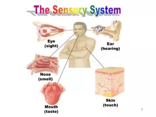
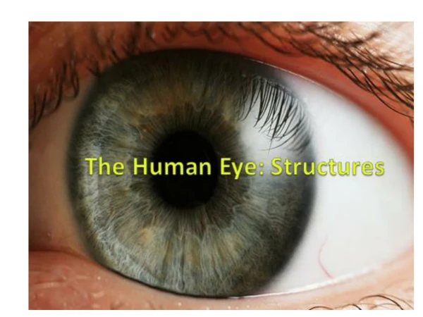
Sensory System structures of the eye. The Five Senses. Eyes Sight Ears Hearing Nose Smell Tongue Taste Skin Touch. Eyes. THE EYE 1” in diameter Protected by: orbital socket eyebrows eyelashes eyelids. Structures of the eyes. External structures Orbit Eyelids

E N D
The Five Senses • Eyes • Sight • Ears • Hearing • Nose • Smell • Tongue • Taste • Skin • Touch 3.03 Remember the structures of the sensory system
Eyes 3.03 Remember the structures of the sensory system
THE EYE • 1” in diameter Protected by: • orbital socket • eyebrows • eyelashes • eyelids
Structures of the eyes • External structures • Orbit • Eyelids • Eyelashes • Conjunctiva • Lacrimal apparatus • Extrinsic muscles 3.03 Remember the structures of the sensory system
Structures of the eyes Let’s look at the external structures
conjunctiva A thin membrane that lines the eyelids and covers part of the eye. It secretes mucus to lubricate our eyes. • Wall of the eye made up of three “coats” http://www.stlukeseye.com/popups/images/Conjunctiva.jpg
SCLERA • Outer layer • White of the eye • Tough coating, helps maintain shape of the eye and protects what’s inside. • Muscles responsible for moving the eyeare attached to the sclera – called EXTRINSIC MUSCLES http://www.cis.rit.edu/people/faculty/montag/vandplite/images/chapter_8/cornea2.jpg
Structures of eyes Extrinsic muscles 3.03 Remember the structures of the sensory system
ANTERIOR CHAMBER filled with AQUEOUS HUMOR, a watery fluid. • POSTERIOR CHAMBER filled with transparent, jellylike substance – VITREOUS HUMOR * *
CORNEA • Front of sclera – clear part (NOblood vessels) • Transparent so light rays can pass through • Gets oxygen and nutrients through our lymph fluid.
CHOROID COAT • Middle layer • Contains blood vessels • Opening in front is the pupil. • Colored, muscular layer surrounding the pupil is called the IRIS. • INTRINSIC MUSCLES – change size of the iris to control amount of light entering the pupil. http://stlukes-eye.com/popups/images/Choroid.jpg
LENS LENS • Crystalline structure located behind the iris and pupil. • Elastic • Disc shaped • biconvex • Situated between the anterior and posterior chambers http://www.visionweb.com/vwweb/images/content/consumers/illustrations/about_the_eye_anatomy_lens2.jpg
RETINA • Innermost layer • Light rays focus an image on the retina • The image travels to the cerebral cortex via the OPTIC NERVE • If light rays don’t focus properly on the retina, corrective lenses can bend the light rays as required. • Retina contains specialized cells called rods and cones http://www.thetech.org/exhibits/online/color/eye/Rods_and.gif
Eyes • Let’s look at the internal structures 3.03 Remember the structures of the sensory system
Structures of the eyes • Rods and cones • Rods • Activate in dim light • Do not perceive color • Cones • Activate in bright light • Perceive color
OPTIC DISC A spot on the retina, known as the “blind spot” – nerve fibers gather here to form the optic nerve. Does not contain any rods or cones.
Cornea Pupil Lens (Where light rays are refracted) Retina Rods & Cones(pick up stimulus) Optic Nerve
Eye Trivia What gives the iris color? Which famous screen actress was noted for her stunning eyes? Many people thought her irises were violet in color… 3.03 Remember the structures of the sensory system
Eyes Name the structures… sclera Ciliary body Ch….. conjunctiva Re…. cornea iris pupil lens Anterior chamber optic nerve Optic d… Extrinsic muscle Review 3.03 Remember the structures of the sensory system
3.04 Functions and disorders of the eye 3.04 Understand the functions and disorders of the sensory system
Understanding the functions of the eye Sight 3.04 Understand the functions and disorders of the sensory system
Understanding the functions of the eye Interesting tidbit External eye • Orbit • Eyelids and eyelashes Women blink twice as often as men. Why do we blink? • Conjunctiva • Lacrimal apparatus - The system that secretes and drains tears into the nasal cavity, consisting of the lacrimal gland, the lacrimal lake, the lacrimal duct, the lacrimal sac, and the nasolacrimal duct. • Extrinsic muscles 3.04 Understand the functions and disorders of the sensory system
The eye is bathed in fluid from our lacrimal glands. The tears empty into our nasal cavity. This is why your nose gets stuffy when you cry!! http://medicalimages.allrefer.com/large/lacrimal-gland-anatomy.jpg
Understanding the functions of the eye Which extrinsic muscle allows you to look upward? External eye Extrinsic muscles are voluntary muscles, external to the eye, that control the direction of the eye movement. 3.04 Understand the functions and disorders of the sensory system
Understanding the functions of the eye Internal eye • Cornea • Iris • Pupil 3.04 Understand the functions and disorders of the sensory system
Understanding the functions of the eye Internal eye • Ciliary body- is the structure in the eye that releases a transparent liquid called the aqueous humor within the eye. • Lens 3.04 Understand the functions and disorders of the sensory system
Understanding the functions of the eye Internal eye • Sclera • Choroid • Retina 3.04 Understand the functions and disorders of the sensory system
Understanding the functions of the eye Internal eye • Vitreous humor = clear gel that fills the space between the lens and the retina of the eyeball 3.04 Understand the functions and disorders of the sensory system
It is a small and highly sensitive part of the retina. It allows us to appreciate detail and perform tasks that require central vision such reading. What is the macula? 3.04 Understand the functions and disorders of the sensory system
Understanding the functions of the eye • Trace the pathway of vision. • Is there anything strange about this picture? Explain 3.04 Understand the functions and disorders of the sensory system
Understanding the functions of the eye Vision What happens as you move your paper away from and toward to your eye? 3.04 Understand the functions and disorders of the sensory system
Check your knowledge! 3.04 Understand the functions and disorders of the sensory system
Disorders of the eye Astigmatism Presbyopia Glaucoma Diabeticretinopathy Detachedretina Hyperopia Color blindness Cataract Myopia Conjunctivitis Have you heard of these conditions? What do you know about them? 3.04 Understand the functions and disorders of the sensory system
Disorders of the eye Cataract Describe this lens. 3.04 Understand the functions and disorders of the sensory system
Disorders of the eye Cataract How is a cataract treated? Initially treated with new glasses or improved lighting. Surgical removal required with progression. 3.04 Understand the functions and disorders of the sensory system
Disorders of the eye Ishihara chart Color blindness Do you see the number? What is color blindness? What causes it? Who is most likely to have color blindness? 3.04 Understand the functions and disorders of the sensory system
Color blindness occurs when there is a problem with the nerve cells of the eye called cones. Most color blindness is due to a genetic problem. About 1 in 10 men have some form of color blindness. Very few women are color blind. 3.04 Understand the functions and disorders of the sensory system
Disorders of the eye How is conjunctivitis spread? How can it be prevented? Conjunctivitis • What is conjunctivitis? • What are the symptoms? • What causes it? • How is it treated? 3.04 Understand the functions and disorders of the sensory system
Conjunctivitis inflammation or infection of the membrane lining the eyelids - the conjunctiva. There are many causes of conjunctivitis. Viruses are the most common cause. "Pink eye" refers to a viral infection of the conjunctiva. These infections are especially contagious among children. Practice good hand washing!!!!!!! Tx = Antibiotic eye drops if bacterial conjunctivitis. Viral conjunctivitis will disappear on its own. Doctors may give a mild antibiotic eye drop for “pink eye” to prevent bacterial conjunctivitis.
Detached retina Retinal detachment often begins when the vitreous gel shrinks and separates from the retina allowing fluid to seep behind the retina causing it to detach. Retinal detachment requires care right away. Without treatment, vision loss can progress from minor to severe or even to blindness within a few hours or days. Surgery is the only way to reattach the retina. In most cases, surgery can restore good vision. 3.04 Understand the functions and disorders of the sensory system
Detached retina Compare this process to the previous picture. What might cause this condition? 3.04 Understand the functions and disorders of the sensory system
Causes of retinal detachment: An eye or head injury leading to tears or holes in the retina. Traction on the retina. Traction pulls the retina away from the layers beneath it. The most common cause of this problem is diabetes. 3.04 Understand the functions and disorders of the sensory system
Disorders of the eye Diabetic retinopathy • What causes diabetic retinopathy? • What are the symptoms? • Explain the impact on vision. 3.04 Understand the functions and disorders of the sensory system
Diabetic retinopathy Diabetes damages the small blood vessels in your retina. This is called diabetic retinopathy. leading cause of blindness in working-age Americans. People with type 1 and type 2 diabetes are at risk for this condition. 3.04 Understand the functions and disorders of the sensory system
What are the symptoms? Most often, diabetic retinopathy has no symptoms until the damage to your eyes is severe. • Blurred vision and slow vision loss over time • Floaters and shadows • Trouble seeing at night 3.04 Understand the functions and disorders of the sensory system
Treatments for diabetic retinopathy Laser eye surgery creates small burns in the retina where there are abnormal blood vessels. A surgical procedure called vitrectomy is used when there is bleeding (hemorrhage) into the eye. Drugs that prevent abnormal blood vessels from growing, and steroid drugs injected into the eyeball are possible new treatments for diabetic retinopathy. 3.04 Understand the functions and disorders of the sensory system
Disorders of the eye Tonometry Glaucoma • What are the common symptoms of glaucoma? • How is it diagnosed? • How is it treated? 3.04 Understand the functions and disorders of the sensory system
Glaucoma Glaucoma refers to a group of eye conditions that lead to damage to the optic nerve. This nerve carries visual information from the eye to the brain. In most cases, damage to the optic nerve is due to increased pressure in the eye, also known as intraocular pressure (IOP). 3.04 Understand the functions and disorders of the sensory system
Causes, incidence, and risk factors Glaucoma is the second most common cause of blindness in the United States. There are four major types of glaucoma: