Download
1 / 15
150 likes | 314 Vues
Chapter 18. The Genetics of Viruses and Bacteria. 0.5 m. Figure 18.1 T4 bacteriophage infecting an E. coli cell. Virus. Bacterium. Animal cell. Animal cell nucleus. 0.25 m. Figure 18.2 Comparing the size of a virus, a bacterium, and an animal cell.

E N D
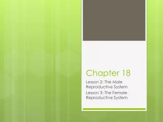
Chapter 18 The Genetics of Viruses and Bacteria
0.5 m Figure 18.1 T4 bacteriophage infecting an E. coli cell
Virus Bacterium Animalcell Animal cell nucleus 0.25 m Figure 18.2 Comparing the size of a virus, a bacterium, and an animal cell
Capsomereof capsid Membranousenvelope RNA Capsomere DNA Head Capsid Tail sheath DNA RNA Tail fiber Glycoprotein Glycoprotein 80 225 nm 18 250 mm 80–200 nm (diameter) 70–90 nm (diameter) 50 nm 20 nm 50 nm 50 nm (d) Bacteriophage T4 (a) Tobacco mosaic virus (b) Adenoviruses (c) Influenza viruses Figure 18.4 Viral structure
VIRUS Entry into cell and uncoating of DNA DNA Capsid Transcription Replication HOST CELL Viral DNA mRNA Viral DNA Capsid proteins Self-assembly of new virus particles and their exit from cell Figure 18.5 A simplified viral reproductive cycle
Attachment. The T4 phage usesits tail fibers to bind to specificreceptor sites on the outer surface of an E. coli cell. Entry of phage DNA and degradation of host DNA.The sheath of the tail contracts,injecting the phage DNA intothe cell and leaving an emptycapsid outside. The cell’sDNA is hydrolyzed. 1 2 4 3 5 Release. The phage directs productionof an enzyme that damages the bacterialcell wall, allowing fluid to enter. The cellswells and finally bursts, releasing 100 to 200 phage particles. Phage assembly Synthesis of viral genomes and proteins. The phage DNAdirects production of phageproteins and copies of the phagegenome by host enzymes, usingcomponents within the cell. Assembly. Three separate sets of proteinsself-assemble to form phage heads, tails,and tail fibers. The phage genome ispackaged inside the capsid as the head forms. Head Tail fibers Tails Figure 18.6 The lytic cycle of phage T4, a virulent phage
Phage DNA The phage attaches to a host cell and injects its DNA. Many cell divisions produce a large population of bacteria infected with the prophage. Phage DNA circularizes Phage Occasionally, a prophage exits the bacterial chromosome, initiating a lytic cycle. Bacterial chromosome Lytic cycle Lysogenic cycle Certain factors determine whether The bacterium reproduces normally, copying the prophage and transmitting it to daughter cells. The cell lyses, releasing phages. Prophage Lytic cycle is induced Lysogenic cycle is entered or New phage DNA and proteins are synthesized and assembled into phages. Phage DNA integrates into the bacterial chromosome,becoming a prophage. Figure 18.7 The lytic and lysogenic cycles of phage , a temperate phage
Glycoproteins on the viral envelope bind to specific receptor molecules(not shown) on the host cell, promoting viral entry into the cell. 1 Capsid RNA Envelope (with glycoproteins) Capsid and viral genome enter cell 2 HOST CELL The viral genome (red) functions as a template forsynthesis of complementary RNA strands (pink) by a viral enzyme. 3 Viral genome (RNA) Template mRNA Complementary RNA strands also function as mRNA, which is translated into both capsid proteins (in the cytosol)and glycoproteins for the viral envelope (in the ER). 5 Capsid proteins New copies of viral genome RNA are made using complementary RNA strands as templates. 4 ER Copy of genome (RNA) Glyco- proteins Vesicles transport envelope glycoproteins to the plasma membrane. 6 New virus 8 7 A capsid assembles around each viral genome molecule. Figure 18.8 The reproductive cycle of an enveloped RNA virus
Glycoprotein Viral envelope Capsid Reversetranscriptase RNA(two identicalstrands) Figure 18.9 The structure of HIV, the retrovirus that causes AIDS
The virus fuses with the cell’s plasma membrane. The capsid proteins are removed, releasing the viral proteins and RNA. 1 HIV Membrane of white blood cell 2 Reverse transcriptase catalyzes the synthesis of a DNA strand complementary to the viral RNA. HOST CELL 3 Reverse transcriptase catalyzes the synthesis ofa second DNA strand complementary to the first. Reverse transcriptase Viral RNA RNA-DNAhybrid 4 The double-stranded DNA is incorporated as a provirus into the cell’s DNA. 0.25 µm HIV entering a cell DNA NUCLEUS Provirus ChromosomalDNA RNA genomefor the nextviral generation 5 Proviral genes are transcribed into RNA molecules, which serve as genomes for the next viral generation and as mRNAs for translation into viral proteins. mRNA 6 The viral proteins include capsid proteins and reverse transcriptase (made in the cytosol) and envelope glycoproteins (made in the ER). 7 Capsids are assembled around viral genomes and reverse transcriptase molecules. 8 Vesicles transport the glycoproteins from the ER to the cell’s plasma membrane. 9 New viruses bud off from the host cell. New HIV leaving a cell Figure 18.10 The reproductive cycle of HIV, a retrovirus
Figure 18.11 SARS (severe acute respiratory syndrome), a recently emerging viral disease (b) The SARS-causing agent is a coronavirus like this one (colorized TEM), so named for the “corona” of glycoprotein spikes protruding from the envelope. (a) Young ballet students in Hong Kong wear face masks to protect themselves from the virus causing SARS.
Originalprion Prion Many prions Normalprotein Newprion Figure 18.13 Model for how prions propagate