Download
1 / 19
190 likes | 371 Vues
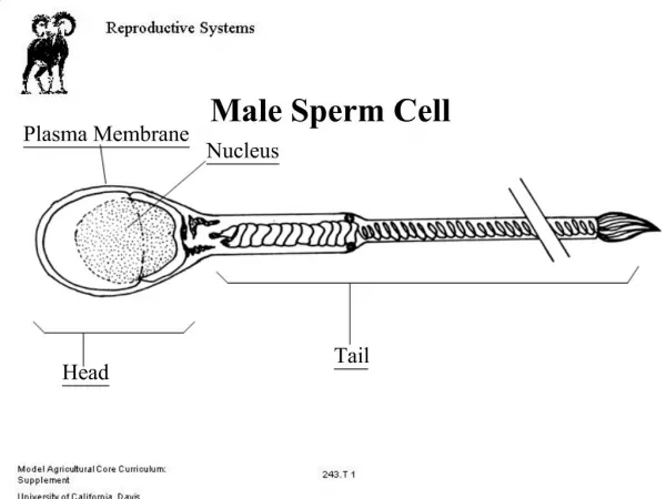
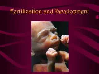
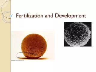
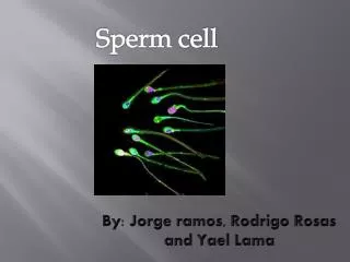
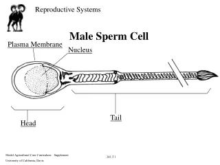
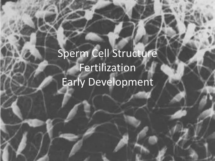
Sperm Cell Structure Fertilization Early Development. Fertilization. Sperm Capacitation. Freshly ejaculated sperm are unable or poorly able to fertilize. They must first undergo a series of changes known collectively as capacitation .

E N D
Sperm Capacitation • Freshly ejaculated sperm are unable or poorly able to fertilize. • They must first undergo a series of changes known collectively as capacitation. • Capacitationis associated with removal of proteins, reorganization of plasma membrane lipids and proteins.
Sperm-ZonaPellucida Binding • Binding of sperm to the zonapellucida is a receptor-ligand interaction • The carbohydrate groups on the zonapellucidaglycoproteins function as sperm receptors. • The sperm molecule that binds this receptor is not known with certainty, and indeed, there may be several proteins that can serve this function.
The Acrosome Reaction • The sperm then faces the daunting task of penetrating the zonapellucida to get to the oocyte. • The acrosome - a huge modified lysosome that is packed with zona-digesting enzymes • The acrosome reaction provides the sperm with an enzymatic drill to get throught the zonapellucida. • Leakage of acrosomal enzymes from the sperm's head. • As the acrosome reaction progresses and the sperm passes through the zonapellucida, more and more of the plasma membrane and acrosomal contents are lost. • Some sperm that lose their acrosomes and are not effective
Penetration of the ZonaPellucida • The force from the sperm's flagellating tail+ acrosomal enzymes, allow the sperm to create a tract through the zonapellucida. • These two factors allow the sperm to traverse the zonapellucida. • Sperm motility is important to zona penetration, allowing the sperm to basically cut its way through the zona
Sperm-Oocyte Binding • Once a sperm penetrates the zonapellucida, it binds to and fuses with the plasma membrane of the oocyte. • The molecular nature of sperm-oocyte binding is not completely resolved. • A leading candidate in some species is a dimeric sperm glycoprotein called fertilin, which binds to a protein in the oocyte plasma membrane and may also induce fusion.
The Zona Reaction • The zona reaction refers to an alteration in the structure of the zonapellucida catalyzed by proteases from cortical granules. • blocks polyspermy in most mammals. • The zonapellucida hardens. • Runner-up sperm that have not finished traversing the zonapellucida by the time the hardening occurs are stopped in their tracks. • Sperm receptors in the zonapellucida are destroyed. • Therefore, any sperm that have not yet bound to the zonapellucida will no longer be able to bind, let alone fertilize the egg.
The one cell embryo undergoes a series of cleavage divisions, progressing through 2-cell, 4-cell, 8-cell and 16 cell stages. A four cell embryo is shown here. The cells in cleavage stage embryos are known as blastomeres.
Early on, cleavage divisions occur quite synchronously. In other words, both blastomeres in a two-cell undergo mitosis and cytokinesis almost simultaneously.
Soon after development of the 8-cell or 16-cell embryo the formation becomes a mass of cells called a morula. • this embryo shown here probably has between 20 and 30 cells. • It is difficult to count the cells in a morula;
Formation of an accumulation of fluid inside the embryo, signals formation of the blastocyst.