Download
1 / 26
290 likes | 553 Vues
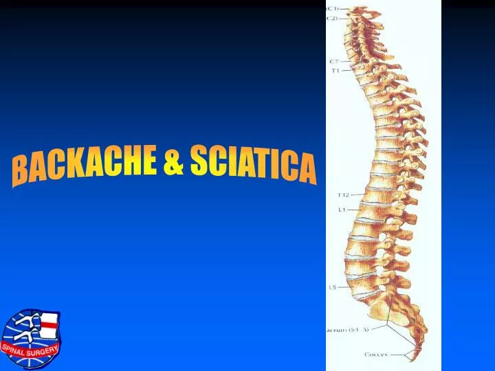
BACKACHE & SCIATICA. LUMBAR SPINE. No. of vertebrae- 5 Lordotic curve- anteriorly Strongest portion of spine Stress- maximum Facet its- sinovial smooth gliding motion restrict motion beyond limits Pedicles vary in lie. Placed more laterally from L1 to L5

E N D
LUMBAR SPINE • No. of vertebrae- 5 • Lordotic curve- anteriorly • Strongest portion of spine • Stress- maximum • Facet its- sinovial • smooth gliding motion • restrict motion beyond limits • Pedicles vary in lie. Placed more laterally from L1 to L5 • L4 and L- Transmit- at. Laterally • maximum mobility
INTERVERTEBRAL DISC • Volume of lumbar discs 10 cm3, • Nuclues 15% = 1.5 cm3, • Annulus has 12 concentric layers very strong • Disc usually is not compressible or very slightly compressible • Intrinsic pressure of muscles = 60 kg/sq cm
THE MUSCLES • The strength of spine lies in its muscles includes paraspinal abdominal quadriceps- power house of the body
BONY CANAL • At L1- Round • At L5- Trifoliate • Lat. Rescusses- prominent at • AP diameter varies from 15 to 25 mm • 20 mm capacious • 12 to 15 mm small canal • 12 mm narrow- spinal stenosis
LIGAMENTUN FLAVUM • Thickness at L5/81 5.5 mm • Thickness at L4/5 6.2 mm • Ligament does not hypertrophy • It buckles in or unfolds
THE MOTION SEGMENT • Motion is produced by structures holding vertebrae together • Intervertebral disc • Intervertebral foramen facets • Interlaminar space • Ligamentum flavum • Inter and spura spinous ligaments A change in the intervertebral disc produces Change in the whole motion segment
MOVEMENTS OF SPINE Mean values in degrees of range of motions (ROM) of the lumbar spine (Punjabi et. al. 1994) Level Flexion-Extension Axial Rotation Lateral Bending L1/2 10.1 2.1 4.9 L2/3 10.8 2.6 7.0 L3/4 11.2 2.6 5.7 L4/5 14.5 2.2 5.7 L5/S1 17.8 1.3 5.5
FREQUENCY OF DISC PROLAPSE Total no. of cases 400 (one level disc prolapse) • Level Percentage • L5/S2 49 • L4/5 40 • L3/4 7.5 • L2/3 3 • L1/2 0.5
CAUSES OF BACKACHE • unusual activity • stressful city life • Occupational • Poor posture • Overweight • PIVD • Facet syndrome • Muscles sprain • Spinal stenosis • Sacroiliac – jt pain • Inflamation trauma, malignancy
REFERRED PAIN • Common in backache • Radiation of pain to areas beyond known dermatomal patterns • Pain relieved by local anaesthetic infiltration in muscles and ligaments • Facet joint pain in produced in buttocks • Source of pain can be other than the disc
FACET SYNDROME • Morning stiffness • Pain in buttocks (referred) • Sitting uncomfortable • Pain from L4 & L5 discs is referred to SI joints)
EXTENSION PAIN • SI joint . • Upper lumbar disc prolapse • Pathology in hip joint
SYMPTOMS • Backpain • Sciatica • Neurogenic claudication • Sensory dysfunction Cold feet Burning Tingling hyparasthesia Saddle pain – genital hypasthesia
BACK SIGNS • Stance- tilted to one side - Movements restricted - Lordosis obliterated - Muscles prominent due to spasm - Tenderness over muscles • Leg: sensations – Hypoasthesia - Reflexes - Weakness of muscles - Wasting in muscles
INVESTIGATIONS • Plain x-ray:- Instability Sacralisation Anatomial abnormally Reduction of disc height • CT scan: shows lateral rescesses better • CT- Myelo- In presence of implants like cardiac, pace makers, ss spinal implants • MRI : Investigation of choice.
DISCOGRAPHY • Injection of a small quantity of contract in disc spacer and production of symptoms • Useful in detecting symptomatic disc in presence of multiple disc • Origin of pain
DIFFERENTIAL DIAGNOSIS • PIVD commonest cause • Spondylolisthesis • Instability • ST joint arthritis • Helps of root • Tuberculuma, granulation tissue • Cysts- saval, synovial • Neuro fibromas • Tumour • Neuropathy- diabetic carcinomatons • Anxiety states- tension from mind slips into the back