Download
1 / 26
260 likes | 344 Vues
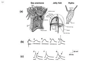
Dermis. Hair shaft. Pore. Dermal papillae (papillary layer of dermis). Epidermis. Meissner's corpuscle. Free nerve ending. Reticular layer of dermis. Sebaceous (oil) gland. Arrector pili muscle. Dermis. Sensory nerve fiber. Eccrine sweat gland. Pacinian corpuscle. Artery.

E N D
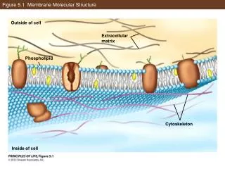
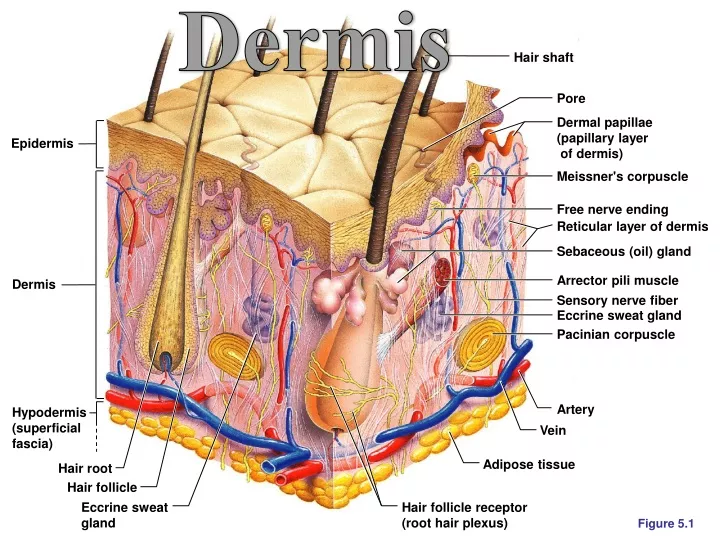
Dermis Hair shaft Pore Dermal papillae (papillary layer of dermis) Epidermis Meissner's corpuscle Free nerve ending Reticular layer of dermis Sebaceous (oil) gland Arrector pili muscle Dermis Sensory nerve fiber Eccrine sweat gland Pacinian corpuscle Artery Hypodermis (superficial fascia) Vein Adipose tissue Hair root Hair follicle Eccrine sweat gland Hair follicle receptor (root hair plexus) Figure 5.1
Dermis • Second major skin region containing strong, flexible connective tissue • Cell types include fibroblasts, macrophages, and occasionally mast cells and white blood cells • Composed of two layers – papillary (bumpy) and reticular (netlike) • Contains a wide variety of accessory organs, including hair follicles, glands, sensory receptors, smooth muscles, and blood vessels
Layers of the Dermis: Papillary Layer • Papillary layer = Stratum Papillarosum • Areolar connective tissue with collagen and elastic fibers • Its superficial surface contains peglike projections called dermal papillae • Dermal papillae contain capillary loops, Meissner’s corpuscles (light touch receptors), and free nerve endings (pain receptors) • Patterns of these bumps found on dermal ridges form fingerprints, which increase friction needed to grip objects. Sweat gland pores along the dermal ridges leave fingerprint patterns on objects we touch.
Layers of the Dermis: Reticular Layer • Reticular layer = Stratum Reticulosum • Accounts for approximately 80% of the thickness of the skin • Named for network of protein fibers criss-crossing throughout the tissue • Collagen fibers in this layer add strength and resiliency to the skin • Elastin fibers provide stretch-recoil properties
Hypodermis • Subcutaneous layer deep to the skin • Composed of adipose and areolar connective tissue
Skin Color • Three pigments contribute to skin color • Melanin – yellow to reddish-brown to black pigment, responsible for dark skin colors. Melanin is formed by melanocytes and secreted; then absorbed by nearby keratinocytes • Different races have different amounts of melanin. • Freckles and pigmented moles – result from local accumulations of melanin • Albinos lack the gene for making melanin. • Carotene – yellow to orange pigment, most obvious in the palms and soles of the feet • Hemoglobin – reddish pigment responsible for the pinkish hue of the skin. The skin color depends on the amount of oxygen carried by the hemoglobin.
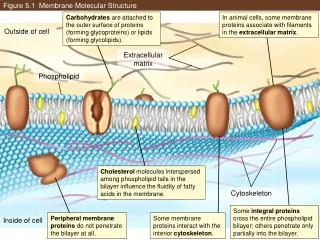
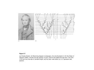
Integumentary Functions Hair shaft Pore Dermal papillae (papillary layer of dermis) Epidermis Meissner's corpuscle Free nerve ending Reticular layer of dermis Sebaceous (oil) gland Arrector pili muscle Dermis Sensory nerve fiber Eccrine sweat gland Pacinian corpuscle Artery Hypodermis (superficial fascia) Vein Adipose tissue Hair root Hair follicle Eccrine sweat gland Hair follicle receptor (root hair plexus) Figure 5.1
Functions of the Integumentary System • Protection • Body temperature regulation • Cutaneous sensation (Touch) • Metabolic functions (Synthesis of molecules) • Water retention • Excretion • (Extras: Homeostasis, Blood Reservoir)
Integumentary Functions: Protection • Skin acts as a chemical, physical, biological, and mechanical barrier • Stratum corneum prevents molecules (including water) to enter or exit • Entire dermis makes physical layer around body • Langerhans cells attack bacteria and viruses
Integumentary Functions: Thermoregulation • Thermoregulation is accomplished by negative feedback interactions between the skin, muscles, and the brain. • Dilation (release heat to environment) and constriction (conserves heat in body) of dermal vessels • Increasing sweat gland secretions to cool the body by evaporation • Activating arrector pili muscles lifts hairs to trap heat and warm the body (more effective in furred mammals than humans) • Hypothalamus of brain is control center (part of midbrain) • Muscles shiver to produce heat when body temperature drops – but this is deeper than the skin
Integumentary Functions: Sense of Touch • Several types of touch receptors embedded in skin • Light touch is sensed by Merkel’s discs (found in epidermis), Meissner’s corpuscles (found in dermal papillae), and root hair plexuses (wrapped around hair follicles). • Deep pressure is sensed by Pacinian corpuscles (found deep in the dermis). • Pain is sensed by free nerve endings throughout the epidermis and dermis. • Cold is sensed by Krause’s corpuscles, found in the dermis. • Heat is sensed by Rufinni’s corpuscles, found in the superficial dermis.
Functions of the Integumentary System • Metabolic functions – synthesis of vitamin D in dermal blood vessels – requires exposure to sunlight • Blood reservoir – skin blood vessels store up to 5% of the body’s blood volume • Excretion – limited amounts of nitrogenous wastes are eliminated from the body in sweat
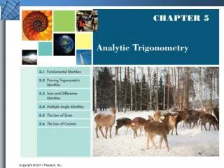
Accessory Organs of the Skin Hair shaft Pore Dermal papillae (papillary layer of dermis) Epidermis Meissner's corpuscle Free nerve ending Reticular layer of dermis Sebaceous (oil) gland Arrector pili muscle Dermis Sensory nerve fiber Eccrine sweat gland Pacinian corpuscle Artery Hypodermis (superficial fascia) Vein Adipose tissue Hair root Hair follicle Eccrine sweat gland Hair follicle receptor (root hair plexus) Figure 5.1
Sweat Glands (Sudoriferous glands) • Different types that either 1. prevent overheating of the body 2. secrete cerumen 3. secrete milk 4. secrete pheromones • Eccrine (merocrine) sweat glands – found all over body, especially in palms, soles of the feet, and forehead • Apocrine sweat glands – found in axillary and anogenital areas – may also release pheromones (molecules that influence the behavior of another organism) • Ceruminous glands – modified apocrine glands in external ear canal that secrete cerumen (“earwax”) • Mammary glands – specialized sweat glands that secrete milk
Sebaceous Glands • Simple alveolar (holocrine) glands found all over the body • Soften skin when stimulated by hormones • Secrete an oily secretion called sebum • Found along hair shafts where the sebum makes its way to the surface by following the hair pore • Blockage of pores causes acne • Sebum also has antibacterial activity
Hair • Filamentous strands of dead keratinized cells produced by hair follicles • Contains hard keratin which is tougher and more durable than soft keratin of the skin • Made up of the shaft projecting from the skin, and the root embedded in the skin • Consists of a core called the medulla, a cortex, and an outermost cuticle
Hair Color • Pigmented by melanocytes at the base of the hair • Color depends on type of pigment released as well as the amount • Dark hair: large amounts of melanin • Blonde hair: intermediate amounts of melanin • Red hair: melanin replaced by trichosiderin • White hair: no melanin or trichosiderin • Gray hair: mix of pigmented & unpigmented hair
Hair Functions • Functions of hair include: • Helping to maintain warmth • Alerting the body to presence of insects on the skin • Guarding the scalp against physical trauma, heat loss, and sunlight • Attracting the opposite sex • Gathering tactile information
Hair Distribution • Hair is distributed over the entire skin surface except: • Palms, soles, and lips • Nipples and portions of the external genitalia
Hair Follicle • Root sheath extending from the epidermal surface into the dermis – includes both epithelial tissue (inner) and connective tissue (outer) regions – boundary between the epi. and conn. tissues is called the glassy membrane (thick basement memb.) • Deep end is expanded forming a hair bulb • A knot of sensory nerve endings (a root hair plexus) wraps around each hair bulb • Bending a hair stimulates these endings, hence our hairs act as sensitive touch receptors
Hair Follicle Longitudinal Section Figure 5.5c
Hair FollicleCross Section Figure 5.5a
Types of Hair • Vellus – pale, fine body hair found in children and the adult female • Terminal – coarse, long hair of eyebrows, scalp, axillary, and pubic regions
Hair Thinning and Baldness • Alopecia – hair thinning in both sexes • True, or frank, baldness • Genetically determined and sex-influenced condition • Male pattern baldness – caused by follicular response to DHT (dihydro testosterone)
Structure of a Nail • Scalelike modification of the epidermis made of keratin found on the distal, dorsal surface of fingers and toes Figure 5.6