Download
1 / 16
170 likes | 449 Vues
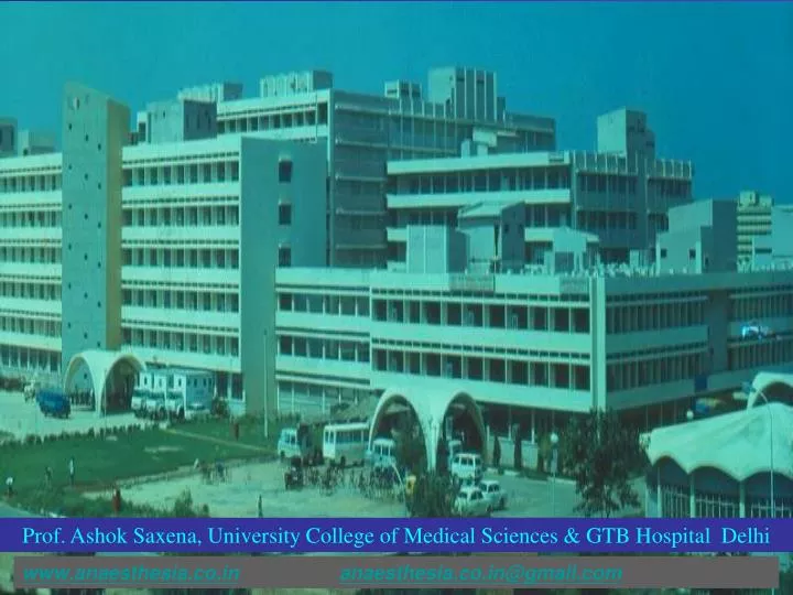
Prof. Ashok Saxena, University College of Medical Sciences & GTB Hospital Delhi. www.anaesthesia.co.in anaesthesia.co.in@gmail.com. Evoked potentials ( E.Ps.).

E N D
Prof. Ashok Saxena, University College of Medical Sciences & GTB Hospital Delhi www.anaesthesia.co.inanaesthesia.co.in@gmail.com
Evoked potentials ( E.Ps.) “E.Ps. are the measurement of the electrical potentials produced in response to stimulating the nervous system (evoked) by sensory, electrical, magnetic or cognitive stimulation” ……Kumar Ashok et al. Anaesthesia 2000; 55: 225-241
E.P. vs EEG • Amplitude of E.Ps. are small compared to EEG : computer averaging used to extract EP signals from the background EEG noise • E.Ps. are : Pathway specific : Stimulus specific : Event-related EPs therefore differ from EEG which is random in nature
Large fiber sensory nerve SSEP Pathway Cell bodies in Dorsal root ganglia Central processes Ipsilateral posterior column Frontoparietal S. M. cortex Ist order neurons synapsing at 3rd order fibers Dorsal column nucleus at cervico-medullary junction Central thalamus 2nd order fibers
SSEP – indications • Carotid endarterectomy** • Descending thoracic aortic surgery** • Spinal embolisation surgery** • Assessing depth of anesthesia • Intropertive monitoring • Evaluation of effects of induction agents, inhal. agents, midazolam etc • Comatose pts, subarach haemg., cereb ischemia, ** - For assessing the integrity of sensory pathway
SSEP recording • Common sites for stimulation : Median N. at wrist, Common peron. N. at knee, Post. tibial. N. at ankle. • Stimulation using electronic stimulator delivering a constant Sq. wave current of 0.1 ms duration, and 3/s in repetition rate : evoked responses picked up by AgCl electrodes on contralateral C3, C4 (2 cm. Post. to vertex and 7 cm. lat.) with ref. to ear electrode. A1, A2 • Filter of 30-300 cycles /sec; Imped : 5000 ohms • Average of 256 evoked responses,
Generators of SSEP after Median Nerve stimulation Peak Generators N9 (EP) Brachial plexus N11 Posterior columns or spinal roots N13/ P13 Dorsal column, Nucleus cuneatus N14/N15 Medial lemniscus N19/P22 Parietal sensory cortex
Effect of anesth. agents on SSEPs • Barbi. , Prop., Etomi. :↑ lat. , ↓ amp. • Ketamine, Midaz. : Normal lat. , ↓ amp. • Fent.,morphine : ↑ lat. , ↓ amp. • Pethidine : ↑ lat. , ↓ amp. • Droperidol , diaz. : ↑ lat. , ↓ amp. • Isofl. , halo. : ↑ lat. , ↓ amp. (Isofl. < 0.4% : No effect)
SSEP monitoring – Spinal surgery • Distraction of spine, placement of pedicle screws & bony decompression : spinal cord itself or N. may be injured • SSEPs moniotrs integrity of posterior aspect of spinal cord : hence, isolated anterior spinal injury may go undetected.
Outcome of SSEP monitoring • 295 pts undergoing spinal stabilisation : neurologic injury rate ↓ed from 6.9% to 0.7% with SSEP monitoring (Meyer et al, 1988) • 100 pts : cervical spine surgery : ↓ in paraplegia from 3.7% to 0% with SSEP monitoring ( Numer et al, 1995)
SSEP during brainstem (post. fossa) & cortical surgery • SSEP monit. to detect cerebral well being to minimize cortical injury from retractions, which occur in 5% of intracranial aneurysms & 10% of cranial base oper. • SSEP to detect cereb. Ischemia in subarachnoid haem. associated with intracranial aneurysm • SSEP utilized during neuroradiology oper, A-V malformations or streptokinase dissolution of occluding blood clots
SSEP monitoring in ICU • Prolongation of CCT in comatose pts. :Worse long term prognosis • CCT ↓ with clinical recovery • Prolongation of CCTin sub arach. Haem. pts associated with transient neurologic deficits • Loss of cortical waves to predict early brain death
Physiol. Factors affecting SSEP • ↓ in MAP to levels below Cereb. Autoregulation : Progressive ↓ in amplitude until loss of waveform ; no change in latency • Hyperthermia - ↓ in amplitude & loss of EP at 42oc • Hypoxia : ↓ in amplitude • Isovol. Hemodilution- progressive ↑ in latency; significant at Hct < 15% ; Ampl. of all waveforms ↓ at Hct < 7 %
SSEP and post-herpetic neuralgia (PHN) • PHN pts : delay in central conduction ↑APL, ↓ amplitude. N19/P22 • Topical application of AAS + diethylethermixture(ADE) tds for 4 wks SSEP recordings : declining trend in APL & ↑ing amplitude of N19/P22 : reflects electrophysiological interaction of antinociceptive effects of topical ADE with generators of SSEP
SSEP and Electroacupuncture • Chronic pain pts. : musculoskeletal origin APL of N19 is significantly ↑ed • Following 1st sitting : APL persists • Following 5th sitting : tends to revert to normal • Following 10th sitting : reverts completely Suggests interaction between EA neural mechanism & thalamic generator of SSEP i.e. N19 ….Kumar Ashok et al, Anaesthesia 1995;50:411-414