Download
1 / 31
310 likes | 314 Vues
Explore the structure and functions of the cerebral cortex, including the motor and sensory areas, association areas, and their role in conscious behavior. Learn about specific areas like Broca's area and the limbic association area.

E N D
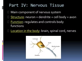
Nervous Part C H. Biology II Adapted 2014-2015
Cerebral Cortex • Thin (2–4 mm) superficial layer of gray matter • 40% of the mass of the brain • Site of conscious mind: awareness, sensory perception, voluntary motor initiation, communication, memory storage, understanding • Each hemisphere connects to contralateral side of the body • There is lateralization of cortical function in the hemispheres
Cerebral Hemispheres • Surface markings • Central sulcus • Separates the precentral gyrus of the frontal lobe and the postcentral gyrus of the parietal lobe • Longitudinal fissure • Separates the two hemispheres • Transverse cerebral fissure • Separates the cerebrum and the cerebellum
Precentral gyrus Central sulcus Postcentral gyrus Frontal lobe Parietal lobe Parieto-occipital sulcus (on medial surface of hemisphere) Lateral sulcus Occipital lobe Temporal lobe Transverse cerebral fissure Cerebellum Pons Medulla oblongata Fissure Spinal cord (a deep sulcus) Gyrus Cortex (gray matter) Sulcus White matter (a) Figure 12.6a
Anterior Longitudinal fissure Frontal lobe Cerebral veins and arteries covered by arachnoid mater Parietal lobe Right cerebral hemisphere Left cerebral hemisphere Occipital lobe Posterior (c) Figure 12.6c
Left cerebral hemisphere Transverse cerebral fissure Brain stem Cerebellum (d) Figure 12.6d
Cerebral hemisphere Diencephalon Cerebellum Brain stem • Midbrain • Pons • Medullaoblongata (d) Birth Figure 12.3d
Functional Areas of the Cerebral Cortex • The three types of functional areas are: • Motor areas—control voluntary movement • Sensory areas—conscious awareness of sensation • Association areas—integrate diverse information • Conscious behavior involves the entire cortex
Motor areas Sensory areas and related association areas Central sulcus Primary motor cortex Primary somatosensory cortex Premotor cortex Somatic sensation Frontal eye field Somatosensory association cortex Broca’s area (outlined by dashes) Gustatory cortex (in insula) Taste Prefrontal cortex Wernicke’s area (outlined by dashes) Working memory for spatial tasks Executive area for task management Primary visual cortex Working memory for object-recall tasks Vision Visual association area Solving complex, multitask problems Auditory association area Hearing Primary auditory cortex (a) Lateral view, left cerebral hemisphere Motor association cortex Primary sensory cortex Primary motor cortex Sensory association cortex Multimodal association cortex Figure 12.8a
Posterior Motor Anterior Motor map in precentral gyrus Toes Jaw Primary motor cortex (precentral gyrus) Tongue Swallowing Figure 12.9
Posterior Sensory Anterior Sensory map in postcentral gyrus Genitals Primary somato- sensory cortex (postcentral gyrus) Intra- abdominal Figure 12.9
Motor areas Sensory areas and related association areas Central sulcus Primary motor cortex Primary somatosensory cortex Premotor cortex Somatic sensation Frontal eye field Somatosensory association cortex Broca’s area (outlined by dashes) Gustatory cortex (in insula) Taste Prefrontal cortex Wernicke’s area (outlined by dashes) Working memory for spatial tasks Executive area for task management Primary visual cortex Working memory for object-recall tasks Vision Visual association area Solving complex, multitask problems Auditory association area Hearing Primary auditory cortex (a) Lateral view, left cerebral hemisphere Motor association cortex Primary sensory cortex Primary motor cortex Sensory association cortex Multimodal association cortex Figure 12.8a
Cingulate gyrus Primary motor cortex Premotor cortex Central sulcus Corpus callosum Primary somatosensory cortex Frontal eye field Parietal lobe Somatosensory association cortex Prefrontal cortex Parieto-occipital sulcus Occipital lobe Processes emotions related to personal and social interactions Visual association area Orbitofrontal cortex Olfactory bulb Primary visual cortex Olfactory tract Fornix Uncus Calcarine sulcus Temporal lobe Primary olfactory cortex Parahippocampal gyrus (b) Parasagittal view, right hemisphere Motor association cortex Primary sensory cortex Primary motor cortex Sensory association cortex Multimodal association cortex Figure 12.8b
Brain Add in PPT • Do some clinical connections/disorders with brain components
Broca’s Area • Anterior to the inferior region of the premotor area • Present in one hemisphere (usually the left) • A motor speech area that directs muscles of the tongue • Is active as one prepares to speak
Anterior Association Area (Prefrontal Cortex) • Most complicated cortical region • Involved with intellect, cognition, recall, and personality • Contains working memory needed for judgment, reasoning, persistence, and conscience • Development depends on feedback from social environment
Limbic Association Area • Part of the limbic system • Provides emotional impact that helps establish memories
Ventricles of the Brain • Connected to one another and to the central canal of the spinal cord • Lined by ependymal cells • Contain cerebrospinal fluid • Two C-shaped lateral ventricles in the cerebral hemispheres • Third ventricle in the diencephalon • Fourth ventricle in the hindbrain, dorsal to the pons, develops from the lumen of the neural tube
Lateral ventricle Septum pellucidum Anterior horn Posterior horn Inferior horn Interventricular foramen Lateral aperture Median aperture Third ventricle Inferior horn Lateral aperture Cerebral aqueduct Fourth ventricle Central canal (a) Anterior view (b) Left lateral view Figure 12.5
Regions and Organization of the CNS • Spinal cord • Central cavity surrounded by a gray matter core • External white matter composed of myelinated fiber tracts
Cortex of gray matter Central cavity Migratory pattern of neurons Inner gray matter Outer white matter Cerebrum Cerebellum Gray matter Region of cerebellum Central cavity Inner gray matter Outer white matter Gray matter Brain stem Central cavity Outer white matter Inner gray matter Spinal cord Figure 12.4
Spinal Cord Trauma • Functional losses • Parasthesias • Sensory loss • Paralysis • Loss of motor function • Flaccid paralysis—severe damage to the ventral root or ventral horn cells • Impulses do not reach muscles; there is no voluntary or involuntary control of muscles • Muscles atrophy
Spinal Cord Trauma • Spastic paralysis—damage to upper motor neurons of the primary motor cortex • Spinal neurons remain intact; muscles are stimulated by reflex activity • No voluntary control of muscles
Spinal Cord Trauma • Transection • Cross sectioning of the spinal cord at any level • Results in total motor and sensory loss in regions inferior to the cut • Paraplegia—transection between T1 and L1 • Quadriplegia—transection in the cervical region
Poliomyelitis • Destruction of the ventral horn motor neurons by the poliovirus • Muscles atrophy • Death may occur due to paralysis of respiratory muscles or cardiac arrest • Survivors often develop postpolio syndrome many years later, as neurons are lost
Amyotrophic Lateral Sclerosis (ALS) • Also called Lou Gehrig’s disease • Involves progressive destruction of ventral horn motor neurons and fibers of the pyramidal tract • Symptoms—loss of the ability to speak, swallow, and breathe • Death typically occurs within five years • Linked to glutamate excitotoxicity, attack by the immune system, or both
Multiple Sclerosis (MS) • An autoimmune disease that mainly affects young adults • Symptoms: visual disturbances, weakness, loss of muscular control, speech disturbances, and urinary incontinence • Myelin sheaths in the CNS become nonfunctional scleroses • Shunting and short-circuiting of nerve impulses occurs • Impulse conduction slows and eventually ceases
Multiple Sclerosis: Treatment • Some immune system–modifying drugs, including interferons and Copazone: • Hold symptoms at bay • Reduce complications • Reduce disability
Developmental Aspects of the CNS • CNS is established during the first month of development • Gender-specific areas appear in both brain and spinal cord, depending on presence or absence of fetal testosterone • Maternal exposure to radiation, drugs (e.g., alcohol and opiates), or infection can harm the developing CNS • Smoking decreases oxygen in the blood, which can lead to neuron death and fetal brain damage
Developmental Aspects of the CNS • Age brings some cognitive declines, but these are not significant in healthy individuals until they reach their 80s • Shrinkage of brain accelerates in old age • Excessive use of alcohol causes signs of senility unrelated to the aging process