Download
1 / 23
230 likes | 519 Vues
Incidental Thyroid Carcinoma Identified by Positron Emission Tomography Scanning Obtained for Metastatic Evaluation. The American Surgeon, Vol 67:p582-584, June 2001 Peyton W. Davis, Nancy D. Perrier, Lee Adler, and Edward A. Levine. Speaker: 8700021 陳修弘. Introduction.

E N D
Incidental Thyroid Carcinoma Identified by Positron Emission Tomography Scanning Obtained for Metastatic Evaluation The American Surgeon, Vol 67:p582-584, June 2001 Peyton W. Davis, Nancy D. Perrier, Lee Adler, and Edward A. Levine. Speaker: 8700021 陳修弘
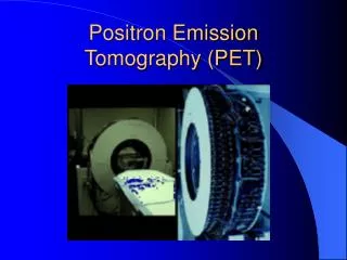
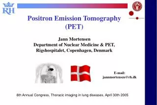
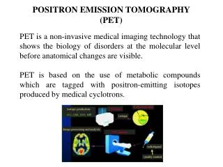
Positron Emission Tomography (PET) ~ I • Tracer: [18F]fluorodeoxyglucose (FDG) • Cells with higher glucose requirements
Positron Emission Tomography (PET) ~ II • Evaluation of patients at risk of having metastatic disease • Staging of malignant melanoma • Therapeutic management of patients with recurrent colorectal carcinoma • Identification of esophageal and gastric carcinoma • Evaluating patients with suspicious thyroid nodules
The Purpose of this Study • To review for cases in which an abnormality was identified by PET in the region or the thyroid
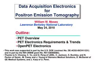
Methods ~ I • Patients were for scanning as part of a metastatic workup • PET scans or the chest or torso for known or suspected extrathyroidal malignancy • 1284 consecutive patients at Wake Forest University Baptist Medical Center • ECAT 951 scanner (CTI, Knoxville, TN) • Between April 1998 and May 2000
Methods ~ II • PET procedure • Patients received a 4-minute-per-bed position transmission scan • Followed by intravenous administration of 15- to 20-mCi FDG • After a 45-min to one-hr delay 7-minute-per-bed position emission imaging was performed
Methods ~ III • The emission scan and the transmission scan were reconstructed and reviewed on a workstation in all three orthogonal planes • The peak activity per cubic centimeter in suspicious areas was corrected for • the dose administered • lean body weight • radioactive decay • blood glucose level to produce a standardized uptake value (SUV).
Methods ~ IV • Four patients with a suspicious area in the thyroid region who were followed up with a fine-needle aspiration (FNA) biopsy under ultrasound guidance. • Two malignant melanoma • One gastric adenocarcinoma • One colon adenocarcinoma
Methods ~ V • They subsequently underwent surgical resection and appropriate adjuvant therapies for their original malignancy • At a later date all cases underwent a definitive resection for the thyroid FNA findings.
Results ~ III • PET scan results: • P1: No other abnormalities • P2: Hilum of the right lung • P3: Gastroesophageal junction and left lung • P4: Suspicious area in the liver • P5: Rectal and thyroid uptake only
Results ~ IV • FNA results • Revealed papillary carcinoma of the thyroid in the five cases • Case five underwent surgery based on uncertainty of a negative ultrasound guided FNA • Histology: supported the diagnosis of papillary carcinoma without any evidence of metastatic disease from the original malignancy. • No patients has suffered a recurrence of the thyroid lesion.
Discussion ~ I • Current methods of detecting thyroid malignancy and recurrence • Sonography • 131I • 123I whole-body scan • Serum thyroglobulin levels • Thallium-201 scintigraphy
Discussion ~ II • PET V.S these other modalities • Superior to scintigraphy in the identification of benign versus malignant thyroid tumors • In one study of six patienst with positive PET and negative 131I scans five had positive histology for carcinoma • PET has a sensitivity of 94% and specificity of 95% fir metastatic disease in patients with negative 131I scan
Discussion ~ III • Other studies have supported the usefulness of PET in evaluating metastatic thyroid disease in patients with negative thallium scans. • FDG PET can stratify these patients into high- and low-risk subsets • FDG PET has definite usefulness in treating patients with known thyroid malignancy or metastases
Discussion ~ IV • In this report, authors address cases in which PET identified an area of occult thyroid carcinoma • These scans were useful in the management of these patients in that metastatic disease was not the cause of their PET abnormality.
Conclusion • These incidental findings suggest • The diagnosis of a primary thyroid malignancy should be entertained for abnormalities of the thyroid observed on PET • Support a potential role for FDG-PET as a component of the evaluation of primary thyroid malignancies