Download
1 / 55
611 likes | 1.04k Vues
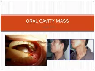
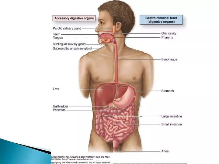
ORAL CAVITY, LIPS AND TONGUE. ORAL CAVITY. The major structures of the oral cavity are the lips , teeth , tongue , oral mucosa and the associated salivary glands. Mastication or chewing is the process by which ingested food is made suitable for swallowing. Chewing involves

E N D
ORAL CAVITY • The major structures of the oral cavity are the lips, teeth, tongue, oral mucosa and the associated salivary glands. • Mastication or chewing is the process by which ingested food is made suitable for swallowing. • Chewing involves • movements of the mandible • cutting and grinding action of the teeth, • activity of the lips and tongue.
The entire oral cavity is lined by a protective mucous membrane, the oral mucosa, which contains many sensory receptors, including the taste receptors of the tongue. • The epithelium of the oral mucosa =stratified squamous keratinized (gum and hard palate). • Non keratinized squamous epithelium (the soft palate, lips, cheeks, and the floor of the mouth).
The oral epithelium is supported by dense collagenous tissue, the lamina propria. • Throughout the oral mucosa, numerous small accessory salivary glands of both serous and mucous types are distributed in the submucosa. • The keratin layer protects the oral mucosa from damage during masticatory function and is best developed on the gingiva (gum) and hard palate.
LIPS Skeletal muscle, connective tissue • SKIN (CUTANEOUS AREA)-SS keratinized epi- sweat and sebaceous glands, hair follicles present • RED AREA (TRANSITION ZONE)-SS non keratinized epi – no hair follicles and glands • ORAL MUCOSA- SS non keratinized. Mucous simple tubular glands: labial salivary glands.
Between the oral portion of the lips and normal skin is the vermilion (V), or the vermilion zone, where epidermis is very thin, lightly keratinized, and transparent to blood in the rich microvasculature of the underlying connective tissue. Because this region lacks the glands for oil and sweat, it is prone to excessive dryness and chapping in cold, dry weather. Internally, the lips contain much-striated muscle (M) and many minor salivary glands
LIPS • The bulk of lips is made up of bundles of circumoral skeletal muscle M seen in transverse section. • The external surface of the lip is covered by hairy skin S which passes through a transition zone to merge with the oral mucosa O of the inner surface. • The transition zone constitutes the free vermilion border of the lip V, and derives its color from the richly vascular dermis, which here has only a thin, lightly keratinized epidermal covering. • .
The free border is highly sensitive due to its rich sensory innervation. • Since the vermilion border is devoid of sweat and sebaceous glands, it requires continuous moistening by saliva to prevent cracking. • The oral mucosa covering the inner surface of the lip has a thick stratified squamous epithelium and the underlying submucosa contains numerous accessory salivary glands G of serous, mucous and mixed seromucous types
CHEEKS • Skeletal muscle • Fibroelastic connective tissue • 1. hairy skin with sweat glands, sebaceous glands. • 2. Muscle core • 3. Oral mucosa. SS non keratinized epithelium. • Submucosa contains elastic fibres
PALATE (SOFT AND HARD) • Hard palate: SS keratinized. • Soft palate: closes off nasopharynx from oropharynx during swallowing. 1. Superior part (nasopharynx): pseudostratified columnar ciliated epi 2. Inferior part (oropharynx): SS non keratinized
supported by a tough, densely collagenous lamina propriaL. • To assist mastication, the palatal mucosa is thrown up into transverse folds or rugae. • The soft palate also has a core of skeletal muscle and lymphoid nodules.
In highly mobile areas such as the soft palate and floor of the mouth, the lamina propria is connected to the underlying muscle by loose submucosal supporting tissue. • In contrast, in areas where the oral mucosa overlies bone, such as the hard palate and tooth-bearing ridges, the lamina propria is tightly bound to the periosteum by a relatively dense fibrous submucosa.
TONGUE - anterior two-thirds • The tongue is a muscular organ covered by oral mucosa (general sensory reception and the special sensory function of taste). The tongue is also vital for speech. • 2 surfaces • upper (dorsal)- rough • lower (ventral)- smooth • A V-shaped groove, the sulcusterminalis, demarcates the anterior two-thirds of the tongue from the posterior one-third. • Epithelium dorsal (SS keratinized) ventral (SS non keratinized)
The body of the tongue consists of a mass of interlacing bundles of skeletal muscle fibresM which permit an extensive range of tongue movements. (longitudinal, transverse and vertical) • The mucous membrane covering the tongue is firmly bound to the underlying muscle by a dense, collagenous lamina propriaLP, which is continuous with the epimysium of the tongue muscle.
LINGUAL PAPILLAE ON DORSAL SURFACE 1. The filiform papillae • The most numerous, • appear as short 'bristles' (pointed) macroscopically. • a heavily keratinized surface projection • consist of a dense supporting tissue core and • Whitish appearance • Lacks taste buds 2. The fungiform papillae • Less numerous • Mushroom shaped, globular • thin non-keratinized epithelium and a • richly vascularized supporting tissue core, • giving them a red appearance macroscopically • Taste buds on their ant surface
3. Circumvallate papillae . • Six to 14 large form a row immediately anterior to the sulcusterminalis • these papillae contain most of the taste buds (only on their lateral sides). • least common type of papillae on the tongue • the largest • encircled by a deep cleft C. • Aggregations of serous glands, called von Ebner's glandsVE, open into the base of the circumvallate clefts, secreting a watery fluid which dissolves food constituents, thus facilitating taste reception. 4. Foliate papillae, which are rudimentary in humans, are found in some animal species.
Numerous small serous and mucous accessory salivary glands are scattered throughout the muscle and lamina propria of the tongue . • SG are stained strongly, whereas the mucous glands MG are poorly stained
The posterior surface of the tongue has a relatively smooth stratified squamous epithelium E overlying lymphoid tissue L containing lymphoid follicles F. • This lymphoid tissue is the lingual tonsil and, with the palatine tonsils and adenoids, completes Waldeyer's ring of lymphoid tissue guarding the entrance to the gastrointestinal and respiratory tracts.
TASTE BUDS • Taste buds are ovoid structures, each containing 50–75 cells, within the stratified epithelium of the tongue and the oral mucosa • About half the cells are elongated gustatory or neuroepithelial cells (taste) cells, which turn over with a 7- to 10-day life span. • Other cells present are slender supportive (sustentacular cells) cells, immature cells, and basal stem cells which divide and give rise to the other two types.
70-80µm long. • 40-50µm wide • The base of each bud rests on the basal lamina and is entered by afferent sensory axons that form synapses on the gustatory cells. • At the apical ends of the gustatory cells microvilli project through an opening called the taste pore. • Molecules (tastants) dissolved in saliva contact the microvilli through the pore and interact with cell surface taste receptors
Taste buds detect at least broad categories of tastants: • metal ions (salty); - tip of tongue • hydrogen ions from acids (sour)- lateral sides • sugars and related organic compounds (sweet); - tip of tongue • alkaloids and certain toxins (bitter)- posterior part of tongue
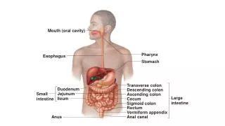
Pharynx • The pharynx, a transitional space between the oral cavity and the respiratory and digestive systems. • The pharynx is lined by stratified non keratinized squamous epithelium in the region continuous with the esophagus and by ciliated pseudostratified columnar epithelium containing goblet cells in the regions close to the nasal cavity.
Esophagus • muscular tube whose function is to transport food from the mouth to the stomach. • It is lined by non keratinized stratified squamous epithelium. • In general, the esophagus has the same major layers as the rest of the digestive tract.
In the lamina propria of the region near the stomach are groups of glands, the esophageal cardiac glands, which also secrete mucus. • In the submucosa are groups of small mucus-secreting glands, the esophageal glands, secretions of which facilitate the transport of foodstuffs and protect the mucosa.
In the proximal third of the esophagus the muscularis is exclusively skeletal muscle like that of the tongue. • The middle third contains a combination of skeletal and smooth muscle fibers • In the distal third the muscularis contains only smooth muscle. • Also, only the most distal portion of the esophagus, in the peritoneal cavity, is covered by serosa. • The rest is enclosed by a layer of loose connective tissue, the adventitia, which blends into the surrounding tissue.