Download

1 / 31
360 likes | 785 Vues
PEPTIC ULCER. Ulcers are defined as a breach in the mucosa of the alimentary tract, which extends through the muscularis mucosa into the submucosa or deeper. ( An erosion differs from an ulcer in being partial thickness mucosal defect).

E N D
Ulcers are defined as a breach in the mucosa of the alimentary tract, which extends through the muscularis mucosa into the submucosa or deeper. ( An erosion differs from an ulcer in being partial thickness mucosal defect). Peptic ulcers are chronic most often solitary, lesions that occur in any portion of gastrointestinal tract exposed to the aggressive action of acid-peptic juices.
Imbalance between aggressive & protective factors Aggressive factors Gastric acid Proteolytic enzyme Protective factors Mucosal layer Bicarbonate secretion Prostaglandins DEFINITION
Gastric physiologyAcid secretion * The human stomach contains about 1 billion parietal cells * These are located in the walls of the midsection of the oxyntic glands * Oxyntic glands are the secretory glands of the gastric mucosa * Oxyntic glands also contin chief, mucous, endocrine, and somatostatin cells
* There are three primary pathways that stimulate gastric acid secretion 1) The neurocrine pathway which delivers transmitters such as acetylcholine post-ganglionic nerves on the stomach wall 2) The endocrine pathway which releases hormones such as gastrin 3) The paracrine pathway which releases histamine * These pathways are interdependent * Once pH is <3.5, pepsinogen is converted to the active proteolytic enzyme pepsin
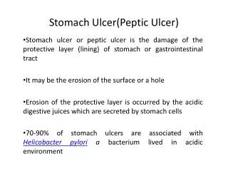
Pathogenesis of peptic ulcer: Peptic ulcers are produced by an imbalance between the gastro-duodenal mucosal defense mechanisms and damaging forces of gastric acid and pepsin, combined with superimposed injury from environmental or immunologic agents.
Imbalance (Aggressive/Defensive Factors) Aggressive Factors * H. pylori * Acid Secretion * Pepsinogen Secretion * NSAIDS * Cigarette smoking * Corticosteroid use
Defensive Factors * Mucus Production * Bicarbonate Production * Mucosal blood flow - more important in the development of stress ulcer * High epithelial cell turnover * Prostaglandins (PGE2) - stimulate mucus and bicarbonate production, and blood flow
Clinical presentation: Remitting, relapsing lesion Most often diagnosed in middle aged to older adults but may first become evident in young adult life. Epigastric burning or aching pain. Pyrosis Pain worse at night and 1 to 3 hours after meal. Nausea, vomiting, bloating , belching and weight loss occur. Complication: Anaemia, hemorrhage, perforation, obstruction. Malignant formation is rare and related to underlying gastritis.
Sites of peptic ulcer: Duodenum: First portion ( few cms from the pyloric ring). Anterior wall is more often affected. Stomach: Usually antrum. Lesser curvature (common) . Anterior and posterior wall and greater curvature (less common). In the margins of a gastroenterostomy (stomal ulcer) In the duodenum, stomach or jejunum of patients with Zollinger-Ellison syndrome. Within or adjacent to a Meckel’s diverticulum that contains ectopic gastric mucosa.
Role ofH. Pylori infection in the pathogenesis of peptic ulcer: H. pylori infection is present in almost all patients with duodenal ulcers and 70% cases with gastric ulcers. Duodenal ulcers - Usually associated with gastritis confined to the antrum. Gastric ulcers - Usually associated with pangastritis. Mechanism: H. pylori secretes urease (generates ammonia), protease (breaks down glycoprotein in the gastric mucus) or phospholipases. Bacterial lipopolysaccharide attracts inflammmatory cells to the mucosa. Neutrophils release myeloperoxide. A bacterial platelet-activating factor promotes thrombotic occlusion of surface capillaries. Mucosal damage allows leakage of tissue nutrients in the surface microenvironment , sustaining the bacillus.
H. Pylori infection in peptic ulceration: (continued) Damage of the protective mucosal layer. The epithelial cells are exposed to the damaging effect of acid-peptic digestion. Inflammation of the gastric mucosa. Chronically inflamed mucosa more susceptible to acid- peptic injury and prone to peptic ulceration. Ulcers occur at sites of chronic inflammation . Eg - Antrum - Junction of antral and body- fundic mucosa (division between the inflamed antral mucosa and normal acid secreting mucosa). Pangastritis - When there is extensive gastritis, the ulcers are more proximally situated. In elderly patients gastric ulcers are more proximally situated as there is proximal migration of the antral-body mucosal junction.
Other factors causing peptic ulcer: Peptic ulcer caused due to high gastrin level and excess acidproduction. Gastrinoma may cause multiple peptic ulceration as in Zollinger Ellison syndrome. There is increased parietal cell mass. Peptic ulcers caused due to impaired mucosal defense . The gastric acid and pepsin levels are normal and no H.pylori are present. Chronic use of NSAIDs (aspirin) causes suppression of mucosal prostaglandin and direct irritative topical effect. Repeated use of corticosteroid in high dose. Cigarette smoking impair healing and favour recurrences. Alcoholic cirrhosis. Personality, psychological stress, ischemia.
Diagnostic Test and Procedures a) Routine * Routine lab tests are not useful in establishing the diagnosis of uncomplicated PUD * Hct, HgB, and stool hemoccult are useful to detect bleeding b) H.pylori test Histology Culture Biopsy/gram stain Biopsy/CLO test Urea breath test Serology
Diagnosis • Breath Test: Carbon 14 Urea Test (see picture) • Blood Test • Endoscopy
Gross features: Gastric ulcers are usually single well delineated lesion. Shape: Round, oval or linear. Size: Usually less than 2cm in diameter. Lesions less than 0.3 cm are likely to be shallow erosions. Giant ulcers are usually greater than 3cm in diameter. May also reach upto 10cm (particularly on lesser curvature ). Mortality rate is higher in these patients. Size does not differentiate benign from malignant ulcer. Some carcinomatous ulcers are less than 4cm and 10% of benign ulcers are more than 4cm .
Gross features: Depth of penetration : Superficial ulcer penetrate the mucosa into the muscularis mucosae. Deeply excavated ulcers having their bases on the muscularis propria. Entire wall is penetrated and the ulcer base is adherant to the pancreas, omental fat or liver. Free perforation into peritoneal cavity may occur.
Biopsy of peptic ulcer: Biopsy is necessary to distinguish between benign and malignant ulcers. Biopsy should be taken from the ulcer edge, at least from each quadrant. Upto 10-12 biopsies may be taken to exclude cancer. Repeat endoscopy may be necessary if biopsies are negative and there is high index of suspicion.
Microscopic features Four distinct layers are present in a peptic ulcer. Surface coat of purulent exudate, bacteria and necrotic debris. Fibrinoid necrosis. Granulation tissue. Fibrosis replacing the muscle wall and extending into subserosa.
Management MEDICAL TREATMENT a) Antisceretory/Anti-acid Agents 1) H2-blockers 2) Proton-pump inhibitors 3) Antacids b) Cytoprotectives 1) Misoprostil 2) Sucralfate c) H. pylori Agents
Eradication of H.pylori (proton pump inhibitor in combination with antibiotics) Cessation of NSAIDS. Criteria for reduction of the size of ulcer crater. Reduction of crater size by 50% over 6-8 weeks of intensive medical management. Endoscopic SURGICAL TREATMENT
Differentials Mesenteric ischemia Angina pectoris Biliary colic Pancreatitis
Complication Hemorrhage Perforation Penetration Obstruction Malignancy