Download
1 / 102
1.22k likes | 2.57k Vues
Mechanical Ventilation. Dr Aidah Abu Elsoud Alkaissi An-Najah National University Faculty of Nursing. Principles of Mechanical Ventilation.

E N D
Mechanical Ventilation Dr Aidah Abu Elsoud Alkaissi An-Najah National University Faculty of Nursing
Principles of Mechanical Ventilation • “… an opening must be attempted in the trunk of the trachea, into which a tube of reed or cane should be put; you will then blow into this, so that the lung may rise again… and the heart becomes • strong…” • --Andreas Vesalius (1555)
Vesalius is credited with the first description of positive-pressure ventilation, but it took 400 years to apply his concept to patient care. • The occasion was the polio epidemic of 1955, when the demand for assisted ventilation outgrew the supply of negative-pressure tank ventilators (known as iron lungs). • In Sweden, all medical schools shut down and medical students worked in 8-hour shifts as human ventilators, manually inflating the lungs of afflicted patients.
Emerson iron lung exemplifying design dating back to the 1930s. Patient's head protrudes through neck collar on left, and electric motor beneath the tank generates negative pressure via the leather bellows on the right. The device weighs 300 kg.
In Boston, the nearby Emerson Company made available a prototype positive-pressure lung inflation device, which was put to use at the Massachusetts General Hospital, and became an instant success. Thus began the era of positive-pressure mechanical ventilation (and the era of intensive care medicine).
Conventional Mechanical Ventilation • The first positive-pressure ventilators were designed to inflate the lungs until a preset pressure was reached. • This type of pressure-cycled ventilation fell out of favor because the inflation volume varied with changes in the mechanical properties of the lungs.
Conventional Mechanical Ventilation • In contrast, volume-cycled ventilation, which inflates the lungs to a predetermined volume, delivers a constant alveolar volume despite changes in the mechanical properties of the lungs. • For this reason, volume-cycled ventilation has become the standard method of positive-pressure mechanical ventilation.
Inflation Pressures • The lungs are inflated at a constant flow rate, and this produces a steady increase in lung volume. • The pressure in the proximal airways (Pprox) shows an abrupt initial rise, followed by a more gradual rise through the remainder of lung inflation. • However, the pressure in the alveoli (PALV) shows only a gradual rise during lung inflation.
Inflation Pressures • The early, abrupt rise in proximal airway pressure is a reflection of flow resistance in the airways. • An increase in airways resistance magnifies (to make greater) the initial rise in proximal airway pressure, while the alveolar pressure at the end of lung inflation remains unchanged.
Inflation Pressures • Thus, when resistance in the airways increases, higher inflation pressures are needed to deliver the inflation volume, but the alveoli are not exposed to the higher inflation pressures. • This is not the case when the distensibility (compliance) of the lungs is reduced.
In this latter condition, there is an increase in both the proximal airways pressure and the alveolar pressure. • Thus, when lung distensibility (compliance) decreases, the higher inflation pressures needed to deliver the inflation volume are transmitted to the alveoli. • The increase in alveolar pressure in noncompliant lungs can lead to pressure-induced lung injury
Cardiac Performance • The influence of positive-pressure ventilation on cardiac performance is complex, and involves changes in preload and afterload for both the right and left sides of the heart.
Cardiac Performance • To describe these changes, it is important to review the influence of intrathoracic pressure on transmural pressure (Pressure gradient across the wall of a blood vessel or organ) , which is the pressure that determines ventricular filling (preload){Preload is the end-diastolic filling pressure of the ventricle just before contraction] and the resistance to ventricular emptying (afterload) {is the force against which the ventricle contracts. A good index of the maximal afterload tension is the peak intraventricularpressure during systole}.
Transmural Pressure • what happens when a normal lung is inflated with 700 mL from a positive-pressure source. • In this situation, the increase in alveolar pressure is completely transmitted into the pulmonary capillaries, and there is no change in transmural pressure (Ptm) across the capillaries.
However, when the same lung inflation occurs in lungs that are not easily distended (panel on the right), the increase in alveolar pressure is not completely transmitted into the capillaries and the transmural pressure increases. • This increase in transmural pressure acts to compress the capillaries.
Therefore, in conditions associated with a decrease in lung compliance (e.g., pulmonary edema, pneumonia), positive-pressure lung inflation tends to compress the heart and intrathoracic blood vessels • This compression can be beneficial or detrimental (damaging, causing harm or injury), as described below.
Preload • Positive-pressure lung inflation can reduce ventricular filling in several ways. • First, positive intrathoracic pressure decreases the pressure gradient for venous inflow into the thorax (although positive-pressure lung inflations also increase intra-abdominal pressure, and this tends to maintain venous inflow into the thorax). • Second, positive pressure exerted on the outer surface of the heart reduces cardiac distensibility, and this can reduce ventricular filling during diastole.
Finally, compression of pulmonary blood vessels can raise pulmonary vascular resistance, and this can impede right ventricular stroke output. • In this situation, the right ventricle dilates and pushes the interventricular septum toward the left ventricle, and this reduces left ventricular chamber size and left ventricular filling. • This phenomenon, known as ventricular interdependence, is one of the mechanisms whereby right heart failure can impair the performance of the left side of the heart.
Afterload • Whereas compression of the heart from positive intrathoracic pressure impedes ventricular filling during diastole, this same compression facilitates ventricular emptying during systole. • This latter effect is easy to visualize (like a hand squeezing the ventricles during systole) and can also be explained in terms of ventricular afterload.
Afterload • That is, ventricular afterload, or the impedance (A measure of the total opposition to current flow in an alternating current circuit),to ventricular emptying, is a function of the peak systolic transmural wall pressure • Incomplete transmission of positive intrathoracic pressure into the ventricular chambers will decrease the transmural pressure across the ventricles during systole, and this decreases ventricular afterload.
Cardiac Output • Positive-pressure lung inflation tends to reduce ventricular filling during diastole but enhances ventricular emptying during systole. • The overall effect of positive-pressure ventilation on cardiac output depends on whether the effect on preload or afterload predominates (To be superior in number, strength, influence, or authority). • When intravascular volume is normal and intrathoracic pressures are not excessive, the effect on afterload reduction predominates, and positive-pressure ventilation increases cardiac stroke output.
The increase in stroke volume causes an increase in systolic blood pressure during lung inflation; a phenomenon known as reverse pulsus paradoxusمتناقض. • The favorable influence of positive intrathoracic pressure on cardiac output is one mechanism that could explain the ability of chest compressions to increase cardiac output during cardiac arrest.
The beneficial actions of positive-pressure ventilation on cardiac output are reversed by hypovolemia. • When intravascular volume is reduced, the predominant effect of positive intrathoracic pressure is to reduce ventricular preload and, in this setting, positive-pressure ventilation decreases cardiac stroke output. • This emphasizes the importance of avoiding hypovolemia in the management of ventilator-dependent patients.
Indications for Mechanical Ventilation • The decision to intubate and initiate mechanical ventilation has always seemed more complicated than it should be. • Instead of presenting the usual list of clinical and physiologic indications for mechanical ventilation, the following simple rules should suffice.
Rule 1. The indication for intubation and mechanical ventilation is thinking of it. • There is a tendency to delay intubation and mechanical ventilation as long as possible in the hopes that it will be unnecessary. • However, elective intubation carries far fewer dangers than emergency intubation, and thus delays in intubation create unnecessary dangers for the patient. • If the patient's condition is severe enough for intubation and mechanical ventilation to be considered, then proceed without delay.
Rule 2. Intubation is not an act of personal weakness. • Housestaff tend to apologize on morning rounds when they have intubated a patient during the evening, almost as though the intubation was an act of weakness on their part. • Quite the contrary, intubation carries the strength of conviction إقناع • (an unshakable belief in something without need for proof or evidence ), and no one will be faulted for gaining control of the airways in an unstable patient.
Rule 3. Initiating mechanical ventilation is not the “kiss of death.” • The perception that “once on a ventilator, always on a ventilator” is a fallacy that should never influence the decision to initiate mechanical ventilation. • Being on a ventilator does not create ventilator dependence; having a severe cardiopulmonary or neuromuscular diseases does.
A New Strategy for Mechanical Ventilation • In the early days of positive-pressure mechanical ventilation, large inflation volumes were recommended to prevent alveolar collapse. • Thus, whereas the tidal volume during spontaneous breathing is normally 5 to 7 mL/kg (ideal body weight), the standard inflation volumes during volume-cycled ventilation have been twice as large, or 10 to 15 mL/kg. .
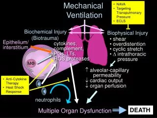
The large inflation volumes used in conventional mechanical ventilation can damage the lungs , and can even promote injury in distant organs through the release of inflammatory cytokines. • The discovery of ventilator-induced lung injury is drastically changing the way that mechanical ventilation is delivered.
Ventilator-Induced Lung Injury • In lung diseases that most often require mechanical ventilation (e.g., acute respiratory distress syndrome [ARDS], pneumonia), the pathologic changes are not uniformly distributed throughout the lungs. • This is even the case for pulmonary conditions like ARDS that appear to be distributed homogeneously throughout the lungs on the chest x-ray. • Because inflation volumes are distributed preferentially to regions of normal lung function, inflation volumes tend to overdistend the normal regions of diseased lungs. • This tendency to overdistend normal lung regions is exaggerated when large inflation volumes are used.
The hyperinflation of normal lung regions during mechanical ventilation can produce stress fractures at the alveolar-capillary interface. • An example a patient with ARDS who required excessively high ventilatory pressures to maintain adequate arterial oxygenation. • These fractures may be the result of excessive alveolar pressures (barotrauma) or excessive alveolar volumes (volutrauma). • Alveolar rupture can have three adverse consequences. • The first is accumulation of alveolar gas in the pulmonary parenchyma (pulmonary interstitial emphysema), mediastinum (pneumomediastinum), or pleural cavity (pneumothorax).
The second adverse consequence is a condition of inflammatory lung injury that is indistinguishable from ARDS • The third and possibly worst consequence is multiorgan injury from release of inflammatory mediators into the bloodstream. This latter process is known as biotrauma.
Modes of Assisted VentilationAssist-Control Ventilation • inflation involves the use of a constant inflation volume instead of a constant inflation pressure. • This method, which is called volume-cycled ventilation, allows the patient to initiate or “trigger” each mechanical breath (assisted ventilation) but can also deliver a preset level of minute ventilation if the patient is unable to trigger the ventilator (controlled ventilation). • This combination is called assist-control ventilation.
Ventilatory Pattern • The tracing begins with a negative-pressure deflection, which is the result of a spontaneous inspiratory effort by the patient. • When the negative pressure reaches a certain level (which is usually set at 22 to 23 cm H2O), a pressure-activated valve in the ventilator opens, and a positive-pressure breath is delivered to the patient.
The second machine breath in the tracing is identical to the first, but it is not preceded by a spontaneous ventilatory effort. The first breath is an example of assisted ventilation, and the second breath is an example of controlled ventilation.
Respiratory Cycle Timing • Volume-cycled ventilation has traditionally employed large inflation volumes (10 to 15 mL/kg or about twice the normal tidal volume during spontaneous breathing). • To allow patients sufficient time to passively exhale these large volumes, the time allowed for exhalation should be at least twice the time allowed for lung inflation.The ratio of inspiratory time to expiratory time, which is called the I:E ratio, should then be maintained at 1:2 or higher.
This is accomplished by using an inspiratory flow rate that is at least twice the expiratory flow rate. • At a normal respiratory rate, an inspiratory flow rate of 60 L/min will inflate the lungs quickly enough to allow the time needed to exhale the inflation volume. • However, when a patient has obstructive lung disease and can't exhale quickly, the I:E ratio can fall below 1:2, and an increase in inspiratory flow rate may be needed to achieve the appropriate I:E ratio.
Work of Breathing • Acute respiratory failure is often accompanied by a marked increase in the work of breathing, and patients who are working hard to breathe are often placed on mechanical ventilation to rest the respiratory muscles and reduce the work of breathing. • However, the assumption that the diaphragm rests during mechanical ventilation is incorrect because the diaphragm is an involuntary muscle that never rests.
The contraction of the diaphragm is dictated by the activity of respiratory neurons in the lower brainstem, and these cells fire automatically and are not silenced by mechanical ventilation. • Only death can silence the brainstem respiratory centers, and the diaphragm follows suit.
This means that the diaphragm does not relax when the ventilator is triggered and delivers the mechanical breath, but it continues to contract throughout inspiration. • Because of the continued contraction of the diaphragm, mechanical ventilation may have little impact on the work of breathing.
Ventilatory Drive • The activity of the diaphragm is largely dictated by the output from the brainstem respiratory neurons, and this output, which is often referred to as the ventilatory drive, is increased as much as three to four times above normal in acute respiratory failure (mechanism unknown). • Reducing ventilatory drive is the appropriate measure for decreasing the workload of the respiratory muscles. • Promoting patient comfort with sedation might help in this regard .
medullary respiratory center: Is composed of several groups of neurons located bilaterally in the medulla oblongata and pons
The Trigger Mechanism • The traditional method of assisted ventilation uses a decrease in airways pressure generated by the patient to open a pressure-sensitive valve and initiate the ventilator breath. • The threshold pressure is usually set at a low level of 21 to 23 cm H2O. • Although this does not seem excessive, many ventilator-dependent patients have positive end-expiratory pressure (PEEP), and this adds to the pressure that must be generated to trigger the ventilator.
For example, if a patient has 15 cm H2O of PEEP and the trigger pressure is 22 cm H2O, a pressure of 7 cm H2O must be generated to trigger a ventilator breath.
This may not seem like much, but the diaphragm generates only 2 to 3 cm H2O during quiet breathing in healthy adults, so generating a pressure of 7 cm H2O will require more than twice the normal effort of the diaphragm.
Disadvantages • Based on an unfounded fear that mechanical ventilation will be accompanied by • progressive atelectasis, large tidal volumes have been employed for volume-cycled ventilation. These volumes are about twice the normal tidal volumes in adults (12 to 15 mL/kg vs. 6 to 8 mL/kg, respectively). • This practice has changed in recent years, and the preferred tidal volumes for mechanical ventilation have been cut in half to the range of 6 • to 8 mL/kg.
Ventilator-Induced Lung Injury • High inflation volumes overdistend alveoli and promote alveolar rupture. • This process is known as volutrauma, and it incites an inflammatory response in the lungs that can produce a condition of inflammatory lung injury similar to the acute respiratory distress syndrome (ARDS). • Inflammatory mediators in the lungs can be released into the systemic circulation and this can lead to inflammatory injury in distant organs. • This condition leads to multiorgan injury
The discovery of volutrauma led to studies comparing ventilation with conventional tidal volumes (12 to 15 mL/kg) and reduced tidal volumes (6 mL/kg). • Some of these studies showed improved outcomes associated with the low-volume lung-protective ventilation • As a result, the recommended inflation volumes for volume-cycled ventilation have • been cut in half to 6 to 8 mL/kg