Download

1 / 21
610 likes | 1.97k Vues
Anatomy of The Kidney. Objectives. By the end of the lecture, the student should be able to describe the: Anatomical features of the kidneys: position, extent, relations, hilum, peritoneal coverings Internal structure of the kidneys: Cortex, medulla and renal sinus

E N D
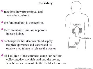
Objectives By the end of the lecture, the student should be able to describe the: • Anatomical features of the kidneys: position, extent, relations, hilum, peritoneal coverings • Internal structure of the kidneys: Cortex, medulla and renal sinus • The vascular segments of the kidneys • The blood supply and lymphatics of the kidneys.
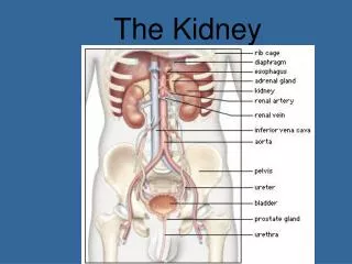
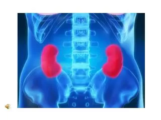
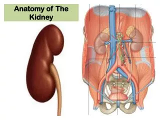
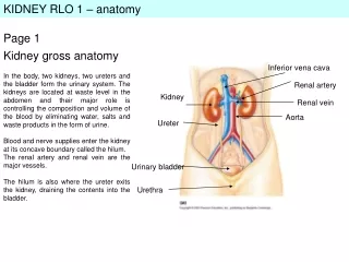
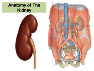
Location & Position of the kidneys • The kidneys are retroperitoneal paired organs • Each kidney lies lateral to the vertebral column, on the posterior abdominal wall largely under cover of the costal margin • In the supine position, the kidneys extend from approximately T12 vertebra superiorly to L3 vertebra inferiorly • The right kidney lies slightly lower than the left kidney because of the large size of the right lobe of the liver. • With contraction of the diaphragm during respiration, both kidneys move downward in a vertical direction by as much as 1 in. (2.5 cm)
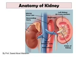
Color, Shape & Dimensions of the kidneys • The Kidneys are reddish brown bean-shaped organs with the dimensions of 12 x 6 x 3cm • Although they are similar in size and shape, the left kidney is a longer and more slender organ than the right kidney, and nearer to the midline. • Each kidneys has: • Convex upper & lower ends • A convex lateral border • A medial border that has a vertical slit called the hilum • Internally the hilum extends into a large cavity called the renal sinus.
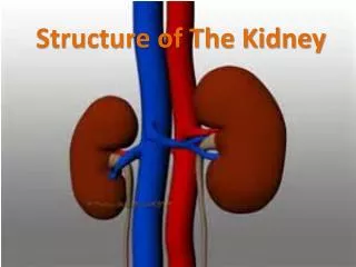
Hilum & Renal sinus • The hilum transmits, from the front backward, the renal vein, renal artery & the ureter (VAU) • Lymph vessels and sympathetic fibers also pass through the hilum. • The renal sinus contains the upper expanded part of the ureter called the Renal pelvis • Perinephric fat continues into the hilum and sinus and surrounds all structures.
Coverings Fibrous capsule: Surrounds the kidney and is closely applied to the outer surface. Perirenal fat: covers the fibrous capsule Renal (Perirenal) fascia: Condensation of connective tissue that lies outside the perirenal fat and encloses the kidney and the suprarenal gland Pararenal fat: Lies external to the renal fascia, is part of the retroperitoneal fat Structures 2,3 & 4 support the kidneys and hold them in position on the posterior abdominal wall.
Relations The posterior surface of the right and left kidneys are related to similar structures The anterior surface of the both kidneys are related to numerous structures, that are different on both sides. some of these structures have an intervening layer of peritoneum and some lie directlyagainst the surface of the kidney.
* *
Relations:Anterior • Left kidney • a small part of the superior pole, on its medial side, is covered by the left suprarenal gland • the rest of the superior pole is covered by the intraperitoneal stomach and spleen • moving inferiorly, the retroperitoneal pancreas covers the middle part of the kidney; • on its lateral side, the lower half of the kidney is covered by the left colic flexure and the beginning of the descending colon, and, • on its medial side, by the parts of the intraperitoneal jejunum.
Relations: Anterior • Right kidney • a small part of the superior pole is covered by the right suprarenal gland • moving inferiorly, a large part of the rest of the upper part of the anterior surface is against the liver and is separated from it by a layer of peritoneum • medially, the descending part of the duodenum is retroperitoneal and contacts the kidney; • the inferior pole of the kidney, on its lateral side, is directly associated with the right colic flexure and, on its medial side, is covered by a segment of the intraperitoneal small intestine.
Relations:Posterior Common to both kidneys • Diaphragm • Costodiaphragmatic recess, of the pleura • Psoas, quadratuslumborum, transversusabdominis muscles • Subcostal (T12), ilio-hypogastric & ilio-inguinal nerves Difference • As the left kidney lies at higher level than the right, it is related to 11th & 12th ribs and the last intercostal space. • The right kidney is related to 12th rib and the last intercostal space.
Vertebrocostal & Renal Angles • Renal angle: The angle between the last rib and the lateral border of erector spinae muscle, is occupied by kidney • Vertebrocostal angle: The angle between the last rib and the lateral border of vertebral column , is occupied by lower part of the pleural sac. Erector spinae Vertebrocostal angle Renal angle
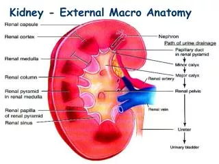
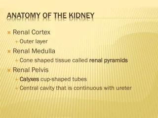
Internal structure • Each kidney consists of an outer renal cortex and an inner renal medulla. • The renal cortex is a continuous band of pale tissue that completely surrounds the renal medulla. • Extensions of the renal cortex, the renal columns project into the inner aspect of the kidney, dividing the renal medulla into discontinuous aggregations of triangular-shaped tissue, the renal pyramids.
The bases of the renal pyramids are directed outward, toward the renal cortex, while the apex of each renal pyramid projects inward, toward the renal sinus. The apical projection (renal papilla) is surrounded by a minor calyx In the renal sinus, several minor calices unite to form a major calyx, and two or three major calices unite to form the renal pelvis, which is the funnel-shaped superior end of the ureters.
Arterial Supply Lobar arteries Interlobar arteries • The renal artery arises from the aorta at the level of the second lumbar vertebra. • Each renal artery divides into 5 segmental arteries that enter the hilum of the kidney, 4 in front of and one behind the renal pelvis. They are distributed to the different segments of the kidney. • Each segmental artery gives rise to number of lobar arteries, each supplies a renal pyramid. • Before entering the renal substance, each lobar artery gives off two or three interlobar arteries Segmental arteries
Arcuate arteries Interlobar arteries Interlobular arteries • The interlobar arteries run toward the cortex on each side of the renal pyramid. • At the junction of the cortex and the medulla, the interlobar arteries give off the arcuate arteries, which arch over the bases of the pyramids. • The arcuate arteries give off several interlobular arteries that ascend in the cortex and give off the afferent glomerular arterioles.
Segmental branches & • vascular segments of kidneys 1 3 5 • Each kidney has 5 segmental branches and is divided into 5 vascular segments: • Apical • Caudal • Anterior Superior • Anterior Inferior • Posterior 4 2 1 3 5 4 2
Blood Supply Glomerulus
Venous Drainage • Both renal veins drain to the inferior vena cava. The left renal vein enters the inferior vena cava a little above the right vein. The left renal vein: • Is three times longer than the right (7.5 cm and 2.5 cm). So, for this reason the left kidney is the preferred side for live donor nephrectomy. • Course: Runsfrom its origin in the renal hilum: • Posterior to the splenic vein and the body of pancreas, and • Then across the anterior aspect of the aorta, just below the origin of the superior mesenteric artery. • Tributaries: • Left gonadal vein enters it from below • Left suprarenal vein, usually receiving one of the left inferior phrenic veins, enters it above but nearer the midline. • The right renal vein: • Lies behind the 2nd part of the duodenum and sometimes the lateral part of the head of the pancreas
Lymphatic Drainage & Nerve Supply Nerve Supply The nerve supply is the renal sympathetic plexus. The afferent fibers that travel through the renal plexus enter the spinal cord in the T10-12 nerves. Lymphatic Drainage The lymph vessels follow the arteries. Lymph drains to the lateral aortic lymph nodes around the origin of the renal artery.