Download
1 / 28
331 likes | 837 Vues
Ultrasonography. The Spleen VCA 341 Dr. LeeAnn Pack lpack@upei.ca. Indications. Splenomegaly Palpable splenic mass Cranial abdominal organomegaly Lethargy, collapse Anemia, abnormal RBC’s. Ultrasound Technique. Left side of body Head of spleen Under border of rib cage on left

E N D
Ultrasonography The Spleen VCA 341 Dr. LeeAnn Pack lpack@upei.ca
Indications • Splenomegaly • Palpable splenic mass • Cranial abdominal organomegaly • Lethargy, collapse • Anemia, abnormal RBC’s
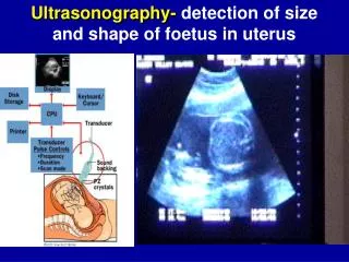
Ultrasound Technique • Left side of body • Head of spleen • Under border of rib cage on left • Body & tail of spleen • Along left body wall • Ventral or lateral to left kidney • Scan sagittal & transverse
Anatomy • Size of normal spleen variable • Assessed subjectively • Enlarged spleen may cross midline or extend caudally to the bladder • Parenchyma • Homogenous, finely textured • Echogenicity • Dog: Spleen > liver > kidney • Cat: Spleen = liver > kidney
Anatomy • Capsule • Smooth, regular, VERY echogenic • Splenic veins • Only other structure normally visualized • Poorly visualized except near hilus • “Whale tail” • Enlargement subjective • Hilus • Check for lymphadenopathy
Pathology • Diffuse splenomegaly • Congestion • Torsion • Inflammation/septicemia • Neoplasia • Lymphosarcoma • Mast cell tumor • Phenothiazine tranquilizers & barbiturate anaesthetics • Extramedullary hematopoesis
Pathology • Focal or multifocal splenic lesions • Hematoma • Infarcts • Cysts • Abscess • Nodular hyperplasia • Neoplasia • Hemangioma • Hemangiosarcoma
Diffuse Splenomegaly • Diffuse increase in echogenicity uncommon • Neoplastic (mast cell or lymphosarcoma) • Diffuse decrease in echogenicity more common • Congestion • Extra-medullary hematopoesis • Lymphosarcoma • Inflammation/ septicemia • Torsion • Normal echogenicity can occur with lymphosarcoma & mast cell tumor
Focal/Multifocal Lesions • More common than diffuse • Anechoic • Cysts • Hematoma/neoplasia • Hypoechoic • Neoplasia • Abscess • Acute infarct • Nodular hyperplasia
Focal/Multifocal Lesions • Hyperechoic • Neoplasia • Abscess • Chronic infarct • Nodular hyperplasia • Mixed echogenicity • Neoplasia • Hematoma • Abscess • Nodular hyperplasia
Torsion • Definitive diagnosis by ultrasound • Characteristic appearance • Severe, diffuse splenomegaly • Hypoechoic • Coarse & “lace-like” • Venous blood flow absent on Doppler • +/- hyperechoic venous thrombi • Lymphosarcoma can appear similar • Normal blood flow
Neoplasia • Lymphosarcoma • Diffuse or focal/multifocal • Hypoechoic or hyperechoic • Can appear normal • Hematoma, hemangioma, hemangiosarcoma • Unable to differentiate • Focal • Hypoechoic, hyperechoic or mixed
Neoplasia • Other neoplasms • Mast cell tumor, leiomyoma, etc. • Presence of peritoneal effusion not a good indication of malignancy • Metastasis • Lungs, liver, lymph nodes (splenic, hepatic, gastric)
Echogenic Focal Lesions • Focal fat deposits • Especially cats • Surround hepatic veins (myelolipomas) • Fibrosis & calcification • Secondary to hematoma, chronic infarcts or granulomas (histoplasmosis) • Primary or metastatic neoplasia
Definitive Diagnosis • Ultrasonic appearance of most splenic diseases non-specific • Consider history, signalment, clinical signs • Fine needle aspirate useful • Biopsy generally not performed
Rupture • Free fluid within the abdomen • Often echoic (due to blood cells) • May be anechoic • Most likely a tumor • Cannot rule out hematoma