Download
1 / 45
460 likes | 705 Vues
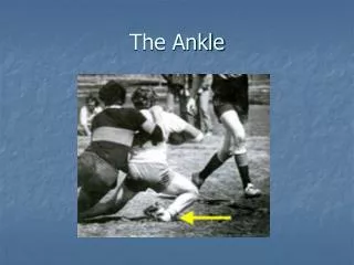
THE ANKLE . By Angela Perkins, Niki Lanier & Justin Longhurst. How can these ankles return to play?. Muscles of the ANKLE. Fibularis ( peroneus ) Longus. Origin : Arises from the head and upper 2/3 of the lateral surface of the fibula.

E N D
THE ANKLE By Angela Perkins, NikiLanier & Justin Longhurst
Fibularis (peroneus) Longus • Origin: Arises from the head and upper 2/3 of the lateral surface of the fibula. • Insertion: This muscle ends in a long tendon that runs behind the lateral mallelous and cross obliquely on the planter surface of the foot to insert at the base of the 1stmetatarsel and medial cuneiform bone. • Action:Everts the foot and is a weak planter flexor of the foot at the ankle.
Fibularis (peroneus) Brevis • Origin: Arises from the distal 2/3 of the lateral surface of the fibula • Insertion: Fibers of this muscle course downward, ending as a tendon that passes behind the lateral malleolus and runs forward to insert into the tuberosity on the lateral side of the base of the 5thmetatarsel • Action: Everts the foot and acts as a weak plantar flexor of the foot at the ankle joint.
Tibialis Anterior • Origin: Arises from the lateral condyle and superior half of the lateral surface of the tibia and from the interosseousmemberane • Insertion: Inserts into the medial and inferior surfaces of the medial cuneiform tarsel and the base of the 1stmetatarsel • Action: Dorsiflexes the foot at the ankle and inverts the foot at the subtalar and midtarsal joints.
Extensor HallucisLongus • Origin: Arises from the middle portion of the anterior surface of the fibula and the interosseous membrane. • Insertion: Inserts on the dorsal aspect of the base of the distal phalanx of the big toe • Action: Extends the big toe, assists in dorsiflexion of the foot at the ankle, and is a weak invertor.
Extensor Digitorum Longus • Origin: Arises from the lateral condyle of the tibia, most of the upper surface of the body of the fibula, and the interosseous membrane. • Insertion: After passing beneath the superior and inferior extensor retinacula, the tendon divides into 4 slips that insert into the middle and distal phalanges of the toes 2 through 5 • Action: Extends the proximal phalanges of the lateral 4 toes and is dorsiflexor of the foot at the ankle.
Gastrocnemius • Origin: This muscle has 2 heads. The lateral head arises from the lateral aspect of the lateral condyle of the femur. The medial head arises from the posterior part of the medial condyle and the popliteal surface of the femur above the medial condyle. • Insertion: The fibers of this muscle unite to form a tendinousraphe. The raphe expands into a broad aponeurosis that unites with the tendon of the soleus and forms the calcaneal tendon. The tendon attaches to the posterior surface of the calcaneus. • Action: Plantar flexes the foot at the ankle, flexes the leg at the knee, and raises the heel during walking.
Soleus • Origin: Arises from the posterior aspect of the head of the fibula, the proximal third of the posterior body of the fibula, the soleal line, and the medial border of the tibia • Insertion: The muscle fibers end in an aponeurosis that thickens, narros, and joins the gastrocnemius. The resulting calcaneal tendon inserts on the posterior surface of the calcaneus • Action: The muscle plantar flexes the foot at the ankle and is an important postural muscle. Apparently, it is constantly active, even during quiet standing, and it aids in maintaining balance.
Plantaris • Origin: Arises from the inferior end of the lateral supracondylar line of the femur and the oblique popiteal ligament. • Insertion: This muscle’s long, slender tendon crosses obliquely between the gastrocnemius and soleus and inserts into the posterior part of the calcaneus, often fusing with the calcaneal tendon. • Action: Weakly assists the gastrocnemius in plantar flexion of the foot at the ankle and flexion of the leg at the knee.
Flexor HallucisLongus • Origin: Arises from the inferior 2/3 of the posterior surface of the fibula and from the inferior portion of the interosseous membrane. • Insertion: Its tendon enters the foot with the tendons of the flexor digitorum longer and the tibialis posterior. It inserts on the base of the distal phalanx of the big toe. • Action: Flexes the distal phalanx of the big toe, planter flexes the foot at the ankle, and helps propel the foot during walking or running.
Tibialis Posterior • Origin: Arises from the posterior surface of the interosseous membrane, the posterior aspect of the tibia inferior to the soleal line, and the posterior surface of the fibula. • Insertion: Inserts on the tuberosity of the navicular bone; the plantar surfaces of the cubiod and cuneiform bones; and the bases of the 2nd, 3rd & 4th metatarsals. • Action: Plantar flexes the foot at the ankle and inverts the foot when the foot is not bearing weight.
Dorsiflexion • Muscles responsible • Tibialis Anterior • Extensor HallucisLongus • Extensor Digitorum Longus • PeroneusTertius • ROM: 20 degrees
Dorsiflexion Stretching • Stretch by forcing ankle into planterflexion • Beginning • Manual force • Performed by patient or clinician • Progressing • Performed by sitting on knees with ankles in plantarflexion • Isolate tibialis anterior by position foot in both plantarflexion & eversion • Isolate toe extensors by positioning foot in both plantarflexion & toe flexion • Isoloateperoneustertitus by postioning foot in both plantarflexion & inversion
Plantar Flexion • Muscle responsible • Achilles Tendon • Tibialis Posterior • Flexor Digitorum Longus • Flexor HallucisLongus • PeroneusLongus • PeroneusBrevis • ROM: 50 degrees
Plantarflexion Stretching • Stretching by forcing ankle into dorsiflexion • Perform with toe straight, toe in & toe out • Beginning • Short-sitting with towel • Manual Pressure • Performed by patient or clinician • Progressing • Standing weight bearing using wall • Incline board • Heel drop off stairs • Isolate gastrocnemius by keeping the knee in extension • Isolate soleus by flexing the knee
Inversion • Muscles responsible • Tibialis Anterior • Tibialis Posterior • Flexor Digitorum Longus • Flexor HallucisLongus • Achilles Tendon • ROM: 5-35 degrees
Inversion Stretching • Stretch by forcing ankle into eversion • Beginning • Manual Pressure • Performed by patient or clinician • Progressing • Performed by rolling ankle into eversion while standing or sitting • Isolate toe flexors by positioning foot into both ankle eversion & toe extension.
Eversion • Muscle responsible • PeroneusBrevis • PeroneusLongus • PeronusTertius • Extensor DigitorumLongus • ROM 5 -15 degrees
Eversion Stretching • Stretch by forcing ankle into inversion • Beginning • Manual pressure • Performed by patient of clinician • Progressing • Performed by rolling ankle into inversion while sitting • Then standing ` • Isolate peroneuslongus & brevis by positioning foot into both inversion & dorsiflexion. • Isolate peroneustertius by positioning foot into both inversion and plantarflexion.
Plantar Fascia Stretching • Any plantarflexor stretch • Massage fascia with a small ball or bottle • Place toes into extension while ankles are dorsiflexed
Beginning Range of Motion Exercises • Ankle Pumps • Leaving the heel on the floor, the patient taps the foot up & down, going as high as possible each time the foot is raised. • Alphabet • Patient spells alphabet with toes leaving heel on the ground. • Toe Exercises • Patient curls, extends, abducts & adducts toes
Toe Exercises • Towel Crunches • Patient is shorting sitting & pulls towel under foot using toes • Progress to wet towel and/or add weight to towel to increase resistances • Begin with 5 towel lengths • Marble pick up (mainly toe flexors) • Patient is short sitting & picks up marble & holds it as long as possible • Progress to heavier objects • Then have patient pick up the objects and move from one container to another. • Begin with 12 marbles
Dorsiflexion StrengtheningIsometric Exercises • Patient is long sitting • Push affected foot into dorsiflexion while resisting with other foot • Don’t move • Hold contraction for 10-15 seconds • Rest 10 second in between • Repeat for 5-10 reps
Dorsiflexion StrengtheningConcentric Exercises • Theraband • Patient is long or short sitting with theraband attached to something anteriorly • Begin with 3 x 8 • Ankle Weights • Patient is long or short sitting with ankle weight strapped to foot • Begin with 3 x 8 • Heel Walking • Patient walks on heels with toes pointed superiorly • Begin with 3 x 30 sec *Isolate tibialis anterior by adding inversion to movement.
Plantarflexion StrengtheningIsometric Exercises • Patient is long sitting, facing a wall • Push affected foot into wall • Don’t move • Patient is short sitting on chair with foot touching the ground • Push affected foot into floor • Don’t move • Hold contraction for 10-15 seconds • Rest 10 second in between • Repeat for 5-10 reps * Isolate gastrocnemius with extended knee, isolate soleus with flexed knee.
Plantarflexion StrengtheningConcentric Exercises • Theraband • Patient is long sitting with therband wrapped around the distal surface of the foot & holding the other end in their hand. • Begin 3 x 8 • Ankle weights • Patient is prone with ankle off the table and ankle weight strapped to foot • Begin with 3 x 8 • Calf Raises • Begin seated • Progress to standing • 2 legs to 1 leg • Standing on a stair with heel hanging off • Holding weights in hands • Leg press machine • Begin with 3 x 8j * Isolate gastrocnemius with extended knee, isolate soleus with flexed knee.
Inversion StrengtheningIsometric Exercises • Patient is sitting adjacent to wall • Push affected foot into wall medially • Don’t move • Patient is short sitting • Push affected foot into inversion while resisting with the other foot • Don’t move • Hold contraction for 10-15 seconds • Rest 10 second in between • Repeat for 5-10 reps
Inversion StrengtheningConcentric Exercises • Theraband • Patient is long or short sitting with theraband wrapped around the distal surface of the foot & holding the other end in their laterally extended ipsilateral hand. • Begin with 3 x 8 • Ankle Weight • Patient is side lying on affected side with ankle weight strapped to foot • Begin with 3 x 8 • Windshield Washers • Patient is short sitting with foot touching ground • Laterally pull towel through full ROM • Progress by wetting towel and/or adding weight to towel • Begin with 5 towel lengths
Eversion StrengtheningIsometric Exercises • Patient is sitting adjacent to wall • Push affected foot into wall laterally • Don’t move • Patient is short sitting • Push affected foot into eversion while resisting with the other foot • Don’t move • Hold contraction for 10-15 seconds • Rest 10 second in between • Repeat for 5-10 reps
Eversion StrengtheningConcentric Exercises • Theraband • Patient is long or short sitting with theraband wrapped around the distal surface of the foot & holding the other end in their laterally extended contralateral hand. • Begin with 3 x 8 • Ankle Weight • Patient is side lying on unaffected side with ankle weight strapped to foot • Begin with 3 x 8 • Windshield Washers • Patient is short sitting with foot touching ground • Medially pull towel through full ROM • Progress by wetting towel and/or adding weight to towel • Begin with 5 towel lengths
Eccentric Exercises • Instead of pulling a joint in the direction of the muscle contraction • Concentric exercises • The muscle decelerates the joint at the end of a movement • Manual resistance
Walking Progression • Non Weight Bearing • Beginning in the deep of the pool • Progress to swallow water • Weight Bearing • Begin on treadmill • Progress to land • Increase speed • Add inclines • Walk backwards
Beginning Proprioception • Two Leg Stance on stable surface • Progress to unstable surface • Mini-tramp, disc, foam rollers, BOSU, wobble/BAPS • Balance Board • Stand on balance board with both legs • Can progress to single leg stance on board • Single Leg Balance • Patient will stand on injured leg while trying not to hold onto anything for support • Progression • Single-leg, arms out to the side, eyes open • Single-leg, arms across the chest, eyes open • Single-leg, arms out to the side, eyes closed • Single-leg, arms across the chest, eyes closed. *Progress by adding movements or perturbations
Advance Proprioception • Single Leg Balance on Disk • Patient will stand on injured leg while balancing on disk • This can also be performed on a half foam roller • Single Leg Wobble Board • Patient centers foot on wobble board & rotates ankle in clockwise & counter-clockwise manner to ensure all sides of board touch the ground • Patient can perform while sitting or to create difficulty can perform standing. • Pro-Fitter • Patient begins with 2 feet & progress to 1 • Use poles, bar, or counter for support • Plyo Toss • Athlete stand on one leg while throwing medicine ball against the rebounder
Speed High movement velocity • Begin with Forward drills • Sprint drills • Straight ladder drills • Proceed to Lateral movement • Box drills • Grapevines • Lateral ladder drills • Proceed to Backward movements
Agility THE ABILITY TO CHANGE DIRECTIONS QUICKLY • Slideboard • Patient begin with end close together &increase distance with progression. • Ladder Drills • Figure 8-Drills • T-Drills • Box Jumping • Patient will stand in the center of the box and jump forward out of the box and backwards back into the box. The patient will then proceed jump in and out of the box in a clockwise & then counter-clockwise direction. Jumping diagnoallly at the corners .
Plyometrics • Vertical jumps • Progress to tuck jump • Progress to depth jumps • Lateral jumps • Start with a small object and progress to larger • Multiple jumps & hops
Cardiovascular Endurance • Minimal or non weight bearing • Pool running with flotation devices • Upper-body ergometer • Rowing machine • Returning to weight bearing • Stationary bike • Elliptical machine • Stairmaster • Returning to running
References • Houglum PA. Therapeutic exercise for musculoskeletal injuries. 2nd ed. Champaign, Il: Human Kinetics; 2005 • Herzog V. Low leg, ankle, and foot rehabilitation In: AT 4201 Febraury 2009; Ogden, UT: 1-36. • Prentice WE. Rehabilitation techniques in sports medicine. 3rd ed. Boston, Ma: WCB/McGraw-Hill; 1999. • Bachle TR , Earle R W. Essentials of strength training and conditioning 3rded. Champaign, Il: Human Kinetics; 2006 • Mattacola CG., Dwyer MK. (2002). Rehabilitation of the ankle after acute sprain or chronic instability. Journal of Athletic training, 37(4):413-429