Download
1 / 66
660 likes | 732 Vues
Chapter 12 – Introduction to the Nervous System. Organization Cell Types. Review. What 3 parts make up the nervous system? Brain Spinal cord Nerves. http://www.nlm.nih.gov/medlineplus/ency/images/ency/fullsize/19588.jpg. Functions of the Nervous System.

E N D
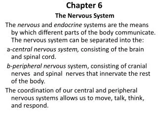
Chapter 12 – Introduction to the Nervous System Organization Cell Types
Review What 3 parts make up the nervous system? • Brain • Spinal cord • Nerves
http://www.nlm.nih.gov/medlineplus/ency/images/ency/fullsize/19588.jpghttp://www.nlm.nih.gov/medlineplus/ency/images/ency/fullsize/19588.jpg
Functions of the Nervous System • Detect changes (stimuli) in the internal or external environment • Evaluate the information • Initiate a change in muscles or glands Goal – maintain homeostasis What does this remind you of??
Organization of the Nervous System • Central nervous system (CNS) • Brain and spinal cord • Peripheral nervous system (PNS) • Nervous tissue in the outer regions of the nervous system • Cranial nerves: originates in the brain • Spinal nerves : originates from the spinal cord • Central fibers: extend from cell body towards the CNS • Peripheral fibers: extend from cell body away from CNS
http://www.nlm.nih.gov/medlineplus/ency/images/ency/fullsize/8679.jpghttp://www.nlm.nih.gov/medlineplus/ency/images/ency/fullsize/8679.jpg
Afferent vs Efferent Nervous pathways are organized into division based on the direction they carry information • Afferent division: incoming information (sensory) • Efferent division: outgoing information (motor) (Efferent = Exit)
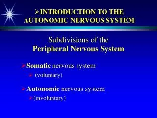
Somatic & Autonomic Nervous Systems Nervous pathways are also organized according to the type of effectors (organs) they regulate • Somatic nervous system (SNS) • Somatic sensory division (afferent) • Somatic motor division (efferent)
Somatic & Autonomic Nervous Systems cont… • Autonomic nervous system (ANS): Carry information to the autonomic or visceral effectors (smooth & cardiac muscles and glands) • Visceral sensory division (afferent) • Efferent pathways • Sympathetic division – “fight or flight” • Parasympathic division – “rest and repair”
http://behavioralphys.wikispaces.com/file/view/autonomic%2520nervous%2520system.gif/162748987/autonomic%2520nervous%2520system.gifhttp://behavioralphys.wikispaces.com/file/view/autonomic%2520nervous%2520system.gif/162748987/autonomic%2520nervous%2520system.gif
Review What are the two main cell types in the nervous system? (Hint: we talked about this when we covered tissue types) Answer: neurons and glia
Cells of the Nervous System Neurons: excitable cells that conduct information Glia (also neuroglia or glial cells): support cells, do not conduct information • Most numerous • Glia = glue
Types of Glia Five major types: • Astrocytes • Microglia • Ependymal cells • Oligodendrocytes • Schwann cells
Astrocytes (12-3A) • Star-shaped, largest, most numerous • Cell extension connect neurons and capillaries • Transfer nutrients from blood to neuron • Help form blood-brain barrier (BBB) http://astrocyte.info/astrocytes1.jpg
Blood-Brain Barrier • Helps maintain stable environment for normal brain function • “feet” of astrocytes wrap around capillaries in brain • Regulates passage of ions • Water, oxygen, CO2, glucose and alcohol pass freely • Important for drug research • Parkinson’s Disease
Microglia (12-3B) • Engulf and destroy cellular debris (phagocytosis) • Enlarge during times of inflammation and degeneration
Ependymal cells (12-3C) • Similar to epithelial cells • Forms thin sheets that line the fluid-filled cavities of the brain and spinal cord • Some cells help produce the fluid that fills these cavities (cerebral spinal fluid - CSF) • Cilia may be present to help circulate fluid http://www.lab.anhb.uwa.edu.au/mb140/corepages/nervous/Images/epen100he.jpg
Oligodendrocytes (12-3D) • Hold nerve fibers together • Produce myelin sheaths in CNS http://4.bp.blogspot.com/_XzEk6ORFLFg/SUQ4IitreiI/AAAAAAAAAD4/XrmtzSv1eGU/s400/article_ms_01.gif http://blustein.tripod.com/Oligodendrocytes/08-zoom.jpg
Multiple Sclerosis (MS) • Most common myelin disorder • Characterized by: • myelin loss and destruction injury and death plaque like lesions • Impaired nerve conduction weakness, loss of coordination, vision and speech problems • Remissions & relapses • Autoimmune or viral infection • Women 20-40 yrs • No known cure
Multiple Sclerosis (MS) http://www.riversideonline.com/source/images/image_popup/ww5r308_big.jpg
Schwann cells (12-3E) • Only in PNS • Support nerve fibers & form myelin sheaths • Satellite cells (12-3G) • Types of schwann cell that covers a neuron’s cell body
http://legacy.owensboro.kctcs.edu/gcaplan/anat/images/Image425.gifhttp://legacy.owensboro.kctcs.edu/gcaplan/anat/images/Image425.gif
Neurons All neurons have 3 parts: • Cell body (soma) • Axon • One or more dendrites
Neuron Anatomy • Soma resembles other cells • Nissl bodies – part of rough ER; contain proteins necessary for nerve signal transmission & nerve regeneration • Dendrites – branch out from soma; receptors; conduct impulse towards soma • Axon – process that extends from the soma at a tapered portion called the axon hillock • Axon collaterals: side branches • Telodendria: distal branches of axon • Synaptic knob: ends of telodendria
http://academic.kellogg.edu/herbrandsonc/bio201_mckinley/f14-3a_structures_in_a__c.jpghttp://academic.kellogg.edu/herbrandsonc/bio201_mckinley/f14-3a_structures_in_a__c.jpg
Neuron Anatomy • Myelin sheaths: areas of insulation produced by Schwann cells; increases speed of nerve impulse • Myelinated = white matter • Unmyelinated = gray matter • Nodes of Ranvier: breaks in myelin sheath btwn Schwann cells • Synapse: junction btwn two neurons or btwn a neuron and an effector
http://academic.kellogg.edu/herbrandsonc/bio201_mckinley/f14-3a_structures_in_a__c.jpghttp://academic.kellogg.edu/herbrandsonc/bio201_mckinley/f14-3a_structures_in_a__c.jpg
Structural Classification of Neurons • Multipolar • One axon, several dendrites • Most numerous • Bipolar • One axon, one dendrite • Least numerous • Retina, inner ear, olfactory pathway • Unipolar • Axon is a single process that branches into a central process (towards CNS) and a peripheral process (towards PNS) • Dendrites at distal end of peripheral process • Always sensory neurons
http://www.google.com/imgres?imgurl=http://psyweb.com/Physiological/Neurons/NImages/Unipolarhttp://www.google.com/imgres?imgurl=http://psyweb.com/Physiological/Neurons/NImages/Unipolar http://www.google.com/imgres?imgurl=http://psyweb.com/Physiological/Neurons/NImages/multipolar http://www.google.com/imgres?imgurl=http://psyweb.com/Physiological/Neurons/NImages/bipolar
Functional Classification of Neurons • Afferent • Sensory • Towards CNS • Efferent • Motor • Towards muscles & glands • Interneurons • Connect afferent & efferent neurons • Lie within CNS
Examples of Reflex Arcs • Ipsilateral • Contralateral • intersegmental
Nerves vs Tracts • Nerves – bundles of parallel neurons held together by fibrous CT in the PNS • Tracts – bundles of parallel neurons in the CNS
Nerve Fibers • Remember the difference between nerves and tracts? • Endoneurium: surrounds each nerve fiber • Perineurium: surrounds fascicles (bundles of nerve fibers • Epineurium: surrounds a complete nerve (PNS) or tract (CNS)
Review: Gray vs White Matter • White matter – myelinated nerve fibers • Myelin sheaths help increase the speed of an action potential • Gray matter – unmyelinated nerve fibers & cell bodies • Ganglia: regions of gray matter in PNS
Nerve Fiber Repair • Nervous tissue has a limited repair capacity b/c mature neurons are incapable of cell division • Repair can take place if soma and neurilemma remain intact
Steps of Nerve Fiber Repair • Injury • Distal axon and myelin sheaths degenerates • Remaining neurilemma & endoneurium forms a “tunnel” from the injury to the effector • Proteins produced in the nissl bodies help extend a new axon down the tunnel to the effector
Nerve Impulses • Neurons are specialized to initiate and conduct signals nerve impulses • Exhibit excitability & conductivity • Nerve impulse wave of electrical fluctuation that travels along the plasma membrane
Membrane Potentials • Difference in charges across the plasma membrane • Inside slightly negative • Outside slightly positive • Result in a difference in electrical charges membrane potential • Stored potential energy • Analogy = water behind a dam
Membrane Potentials • Membrane potential creates a polarized membrane • Membrane has – pole & + pole • Potential difference of a polarized membrane is measured in millivolts (mV) • The sign indicates the charge of the inside of a polarized membrane
Resting Membrane Potential (RMP) • When not conducting electrical signals, a membrane is “resting” • -70mV • RMP maintained by ionic imbalance across membrane • Sodium-Potassium Pump • Pumps 3 Na+ out for every 2 K+ pumps in • Creates an electrical gradient (more positive on outside)
Local Potential • Local potential - The slight shift away from the RMP • Isolated to a particular region of the plasma membrane • Stimulus-gated Na+ channels open Na+ enters membrane potential to moves closer to zero (depolarization) • Stimulus-gated K+ channels open K+ exits membrane potential away from zero (hyperpolarization) • **Local potentials do not spread to the end of the axon**
Action Potentials Definitions: • Membrane potential of an active neuron (one that is conducting an impulse • Action potential = nerve impulse • An electrical fluctuation that travels along the plasma membrane
Steps of Producing an Action Potential (table 12-1) • A stimulus triggers stimulus-gated Na+ channels to open Na+ diffuses inside the cell depolarization • Threshold potential is reached (-59mV) voltage-gated Na+ channels open depolarization continues • Action potential peaks at +30mV, voltage-gated Na+ channels close • Voltage-gated K+ channels open K+ diffuses outward repolarization • Brief period of hyperpolarization (below -70mV) RMP is restored by Na+/K+ pump