Download
1 / 18
190 likes | 394 Vues
ECG Analysis using Wavelet Transforms. By Narayanan Raman Vijay Mahalingam Subra Ganesan Oakland University, Rochester. Objectives. ECG background Wavelet transforms Proposed schemes Conclusion. Electrocardiograph. Electrical activity of the heart, condition of the heart muscle.

E N D
ECG Analysis using Wavelet Transforms By Narayanan Raman Vijay Mahalingam Subra Ganesan Oakland University, Rochester
Objectives • ECG background • Wavelet transforms • Proposed schemes • Conclusion
Electrocardiograph • Electrical activity of the heart, condition of the heart muscle. • Waves are inscribed on ECG during myocardial depolarization and repolarization. • Usually time-domain ECG signals are used. • New computerized ECG recorders utilize frequency information to detect pathological condition.
Electrocardiograph • ECG consists of P-wave, QRS-complex, the T-wave and U-wave. • P-wave-depolarization of atria. • QRS-complex-depolarization of ventricles. • T-wave-repolarization of ventricles. • Repolarization of the atria not visible. • QRS complex detection-most important task in automatic ECG analysis.
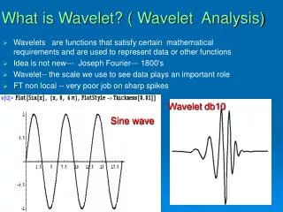
Why wavelet transform? • ECG signal-sequence of cardiac cycles or ‘beats’. • ECG is not strictly a periodic signal-differences in period and amplitude level of beats. • Each region has different frequency components-QRS has high frequency oscillations,T region has lower frequencies,P and U regions have very low frequencies. • Signal contains noise components due to various sources that are suppressed during processing of ECG signal.
Why wavelet transform? (contd.) • Fourier Transform - provides only frequency information, time information is lost. • Short Term Fourier Transform (STFT) - provides both time and frequency information, but resolves all frequencies equally. • Wavelet transform - provides good time resolution and poor frequency resolution at high frequencies and good frequency resolution and poor time resolution at low frequencies. • Useful approach when signal at hand has high frequency components for short duration and low frequency components for long duration as in ECG.
Discrete Wavelet Transform (DWT) • Time-scale representation of signal obtained using digital filtering techniques. • Resolution of the signal is changed by filtering operations. • Scale is changed by upsampling and downsampling (subsampling) operations. • Subsampling-reducing sampling rate, or removing some of the samples of the signal. • Upsampling-increasing sampling rate by adding new samples to the signal.
DWT Analysis • DWT of original signal is obtained by concatenating all coefficients starting from the last level of decomposition. • DWT will have same number of coefficients as original signal. • Frequencies most prominent (appear as high amplitudes) are retained and others are discarded without loss of information.
Proposed Scheme • QRS detection-delineate individual beats in ECG signal. • Real time algorithm-includes noise filtering and use of adaptive thresholds for reliable detection. • Signal is passed through a digital bandpass filter (5 to 15 Hz)-by cascading a low and a high pass filter. • Passes high frequency components of QRS region and suppresses noise and medium frequency T waves. • Filtering of noise and T waves permits use of lower thresholds leading to increased sensitivity of beat detection. • Filter designs use integer coefficients, resulting in faster computations.
Proposed Scheme (contd.) • Transfer functions and corresponding differential equations of filters are defined. • Large slopes of QRS used-slope information obtained by passing signal through a differentiator (high pass filter). • Slope information enhanced by squaring the differentiator output. • Selective amplification of QRS and noise spikes in passband. • Squared o/p passed through moving window integrator. • Output of integrator-large amplitude pulse for every QRS, lower amplitudes for noise spikes.
Proposed Scheme (contd.) • Comparing this pulse amplitude with a suitable threshold, QRS peak is identified. • Adaptive threshold is used-value is continuously updated. • If filtered ECG and integrator output exceed their thresholds, peak is classified as QRS peak. • Monitored by computing estimate of signal level and threshold.
Period and Amplitude Normalization • Normalization eliminates period and amplitude level differences-improves correlation across beats. • Amplitude normalization-dividing sampled values of each beat by the value of the largest peak in that beat. • Period normalization-converting variable length beats into beats of fixed length. • Apply DCT to each beat signal to obtain transform of the same length. • Append zeroes to transform domain signal so that resulting signal length equals normalized length. • Apply inverse transform on this signal to get normalized time domain beat signal.
Wavelet Transform • Each region of oscillations in a beat-wavelets localized at that region. • Amplitudes, time shifts and scale factors of a few wavelets need to be stored. • Mallet pyramidal (sub-band coded) DWT algorithm is used. • Involves 4 stages of complementaryfilter pairs, each stage followed by a downsampler. • Downsampling is by factor of 2-hence number of samples need to be a power of 2.
Conclusions • ECG of normal heart. • ECG of afflicted heart. • QRS peaks identified. • Analysis being done.