Download
1 / 69
690 likes | 814 Vues
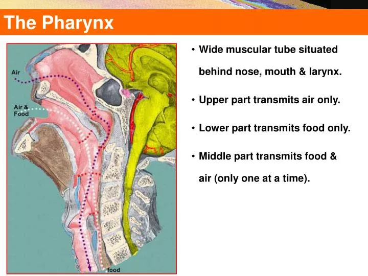
The Pharynx. Wide muscular tube situated behind nose, mouth & larynx . Upper part transmits air only . Lower part transmits food only . Middle part transmits food & air (only one at a time). The Pharynx. Wide muscular tube situated behind nose, mouth & larynx Upper part transmits air only

E N D
The Pharynx • Wide muscular tube situated behind nose, mouth & larynx. • Upper part transmits air only. • Lower part transmits food only. • Middle part transmits food & air (only one at a time).
The Pharynx • Wide muscular tube situated behind nose, mouth & larynx • Upper part transmits air only • Lower part transmits food only • Middle part transmits food & air (only one at a time)
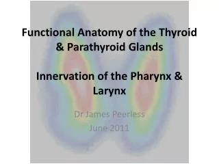
The Pharynx • Muscular tube lying behind the nose, oral cavity & larynx • Extends from the base of the skull to level of the 6th cervical vertebra, where it is continuous with the esophagus • The anterior wall is deficient and shows (from above downward): • Posterior nasal apertures • Opening of the oral cavity • Laryngeal inlet
The PharyngealWall • It is a musculo-membranous wall, composed of: • Mucosa & submucosa • Pharyngobasilar fascia • Muscles: circular & longitudinal • Buccopharyngeal fascia • The buccopharyngeal fascia is separated from the prevertebral fascia by the retropharyngeal space.
The Pharyngeal Facia Bacterial Infection all the way into the abdomen Alar Fascia 1 1 -- Retropharyngeal Space Between Buccopharyngeal and Alar Fascia Buccopharyngeal Fascia Esophagus 2 -- Danger Zone Between Alar and Prevertebral Fascia Trachea Prevertebral Layer 2 Pretracheal fascia includes alar and buccopharyngeal Pericardium
The Pharynx Important Landmarks Nasal Cavity Nasopharynx • Soft Palate • Epiglottis • Cricoid Cartilage Hard palate 1 Oral Cavity Oropharynx 2 Larygopharynx 3 Larynx Trachea
Pharynx Nasopharynx Laryngopharynx Choanae Pharyngeal isthmus Oropharynx Faucial isthmus Epiglottis Eosophagus Trachea Larynx
SYSTEMA RESPIRATORIUM Nasal Part • Boundaries: • Roof:Body of sphenoid & basal part of the occipital bone. • Floor:Upper surface of soft palate & the pharyngeal isthmus (opening between the free margin of soft palate and posterior pharyngeal wall)
SYSTEMA RESPIRATORIUM Nasal Part FUNCTIONS • Respiratory- no food ever enters it. • Walls are rigid & non-collapsible so air passage is kept patent. • Lined by ciliated columnar epithelium • Mucus membrane supplied by trigeminal nerve.
SYSTEMA RESPIRATORIUM Nasal Part • Nasopharynx • Choanae and pharyngeal isthmus • Pharyngeal ostium of the auditory • tube • Auditory tube = salpinx = Eustachian • tube • Torus levatorius • Torus tubarius C2
Nasal Part • Nasopharynx • Choanae and pharyngeal isthmus • Pharyngeal ostium of the auditory • tube • Auditory tube = salpinx = Eustachian • tube • Torus levatorius • Torus tubarius C2
Nasal Part • Tubal tonsil (Gerlach’s tonsil) • Salpingopalatine fold • Salpingopharyngeal fold • Pharyngeal recess • (Rosenmüller‘s fossa) • Fornix • Pharyngeal tonsil (adenoids) C2
Nasal Part • Tubal tonsil (Gerlach’s tonsil) • Salpingopalatine fold • Salpingopharyngeal fold • Pharyngeal recess • (Rosenmüller‘s fossa) • Fornix • Pharyngeal tonsil (adenoids) C2
Nasal Part NASOPHARYNX Sensory CN V2 Torus tubarius Muscles of Soft Palate 1 - Levator veli palatini – CN X 2 - Muscularis uvulae – CN X 3 -Tensor veli palatini – CN V3 1 3 1 Trigeminal / Semilunar ganglion 2 Tensor – mandibular n. 2 1 Muscles of Pharynx 1 – Salpingopharyngeus – CN X 2 – Palatopharyngeus – CN X 2 Pterygoid hamulus of the Medial Pterygoid Plate
SYSTEMA RESPIRATORIUM Oral Part • Lies behind the mouth • Extends from soft palate to upper border of epiglottis • Boundaries: • Roof: soft palate and pharyngeal isthmus • Floor: posterior one third of tongue, median & lateral glossoepiglottic folds, and the valleculae
SYSTEMA RESPIRATORIUM Oral Part • Middle part situated behind oral cavity. • Walls made of superior, middle, inferior constrictors. • Communications • Above : nasopharynx via pharyngeal isthmus • In front: oropharynx via oropharyngeal isthmus • Behind: Supported by C2 + C3 vertebrae • Lateral wall: Palatine tonsil lying in tonsillarfossa(bounded by palatoglossal & palatopharyngeal arches)
Oral Part • Oropharynx: • Soft palate–hyoid bone • Fauces (throat) • Faucial isthmus • Palatoglossal arch • Palatopharyngeal arch • Tonsillar fossa • Palatine tonsil • Semilunar fold • Supratonsillar fossa C2 C3
Oral Part OROPHARYNX Sensory – CN IX Vallecula Palatopharyngeus m. CN X Palatine Tonsils Palatoglossus m. CN X Named were it inserts Gag Reflex Afferent CN IX Efferent CN X
Laryngeal Part Lowest part situated behind larynx • Extends from upper border of epiglottis to lower border of cricoid cartilage • ANTERIOR WALL • Inlet of larynx • Cricoid & arytenoid cartilages • POSTERIOR WALL • C3-6 vertebrae • LATERAL WALL • Has a depression- piriform fossa
Clinical Notes • Adenoides (enlarged pharyngeal tonsils) & adenoidectomy. Adenoids results in obstruction to nasal breathing and make mouth breathing necessary. The patient develops a typical facial expression called the ‘adenoid facies’. May also cause impaired hearing • Otitis media (middle ear infection), secondary to infection of nasopharynx • Tonsillitis & Tonsillectomy Adenoid facies
Clinical Notes • Peritonsillarabcess: complication of tonsillitis and consists of a collection of pus beside the tonsil (peritonsillar space). • Piriformfossa: a common site for the lodging of foreign bodies • Pharyngeal pouch:posteromedialherniation of mucosal diverticulum between thyropharyngeal and cricopharyngeal parts of the inferior constrictor muscle leading to dysphagia (difficulty in swallowing) . It occurs mainly in older people • Retropharyngeal abcess: may spread to the superior mediastinum
Muscles of Pharynx 3 CONSTRICTORS Superior Middle Inferior 3 LONGITUDINAL MUSCLES Stylopharyngeus Salpingopharyngeus Palatopharyngeus
Circular (Constrictor) Muscles • Three in number: Superior, Middle & Inferior • Extend around the pharynx and are inserted posteriorly into a fibrous raphe that extends from the pharyngeal tubercle on the occipital bone to the esophagus • The three muscles overlap each other • The gap between the superior border of the superior constrictor and the occipital bone is filled by thickened pharyngobasilar fascia
Circular (Constrictor) Muscles • Superior constrictor • Origin: medial pterygoid plate, pterygoidhamulus, pterygomandibular ligament, mylohyoid line • Insertion: pharyngeal tubercle, pharyngeal raphe • Middle constrictor • Origin: lower part of stylohyoid ligament, greater & lesser cornu of hyoid bone • Insertion: pharyngeal raphe
Circular (Constrictor) Muscles • Inferior constrictor • Origin: lamina of thyroid cartilage, cricoid cartilage • Insertion: pharyngeal raphe • Functions: • The constrictor muscles propel the bolus of food down into the esophagus • Cricopharyngeus(lower fibers of the inferior constrictor) act as a sphincter, preventing the entry of air into the esophagus between the acts of swallowing
Longitudinal Muscles • Three in number: • Stylopharyngeus • Salpingopharyngeus • Palatpharyngeous • Function: • Elevate the larynx & pharynx during swallowing
Longitudinal Muscles • Stylopharyngeus • Origin:styloid process • Insertion:posterior border of thyroid cartilage • Salpingopharyngeus • Origin:auditory tube • Insertion: blends with palatoglossus • Palatopharyngeus • Origin:palatine aponeurosis • Insertion:posterior border of thyroid cartilage
Nerve Supply to Pharynx • Sensory Nerve Supply: • Nasopharynx: Maxillary nerve • Oropharynx: Glossopharyngeal nerve • Laryngopharynx:Internal laryngeal branch of the vagus nerve • Motor Nerve Supply: • All the muscles of pharynx, except the stylopharyngeus, supplied by the pharyngeal plexus • The stylopharyngeus is supplied by the glossopharyngeal nerve
Blood Supply & Lymphatics • Arterial supply is derived from branches of: • Ascending pharyngeal artery • Ascending palatine artery • Facial artery • Maxillary artery • Lingual artery
Blood Supply & Lymphatics • Arterial supply is derived from branches of: • Ascending pharyngeal artery • Ascending palatine artery • Facial artery • Maxillary artery • Lingual artery
Blood Supply & Lymphatics • Branches from: • External carotid artery • Facial artery • Lingual artery • Branches of maxillary artery
Blood Supply & Lymphatics • Venous supply The veins drain into pharyngeal venous plexus, which drains into the internal jugular vein
Blood Supply & Lymphatics VENOUS DRAINAGE Plexus on posterolateral aspect of pharynx Drain into IJV & facial vein
Blood Supply & Lymphatics • The lymphatics drain into the deep cervical lymph nodes either directly, or indirectly via the retropharyngeal or paratracheal lymph nodes LYMPHATIC DRAINAGE
TheEsophagus • Anatomy of the Esophagus • Muscular tube about 25 cm long • Lined by stratified squamous epithelium • Posterior to trachea • Penetrates diaphragm at esophageal hiatus • Possess upper and lower esophageal sphincters • Sphincter—A circular band of muscle that can pinch close a muscular tube
TheEsophagus • Secretes mucous, transports food – no enzymes produced, no absorption • Mucosa – protection against wear and tear • Submucosa • Muscularis divided in thirds • Superior 1/3 skeletal muscle • Middle 1/3 skeletal and smooth muscle • Inferior 1/3 smooth muscle • 2 sphincters – upper esophageal sphincter (UES) regulates movement into esophagus, lower esophageal sphincter (LES) regulates movement into stomach • Adventitia – no serosa – attaches to surroundings
TheEsophagus • A muscular tube; 25 cm in length • Collapsed at rest, • Flat in upper 2/3 & rounded in lower 1/3 • Commences at the lower border of the cricoid cartilage.(C6). • Descends along the front of the spine, through the posterior mediastinum, passes through the diaphragm, and, entering the abdomen, terminates at the cardiac orifice of the stomach, opposite the eleventh dorsal vertebra. • In the newborn upper limit at the level of 4th or 5th cervertb and it ends at 9th dorsal
TheEsophagus • General direction of the esophagus • is vertical. • Presents two or three slight curvatures. • At commencement, in the median line • Inclines to the left side at the root of the neck • Gradually passes to the middle line • Again deviates to the left
TheEsophagus • The oesophagus also presents an antero-posterior flexure, corresponding to the curvature of the cervical and thoracic portions of the spine. • It is the narrowest part of the alimentary canal, being most contracted at its commencement, and at the point where it passes through the diaphragm.
TheEsophagus • In the neck, the oesophagus is in relation, • in front, with the trachea; and, at the lower part of the neck, where it projects to the left side, with the thyroid gland and thoracic duct; • behind, it rests upon the vertebral column and Longuscolli muscle; on each side, it is in relation with the common carotid artery (especially the left, as it inclines to that side), and part of the lateral lobes of the thyroid gland; the recurrent laryngeal nerves ascend between it and the trachea.
TheEsophagus • In the thorax, it is at first situated a little to the left of the median line: it passes across the left side of the transverse part of the aortic arch, descends in the posterior mediastinum, along the right side of the aorta, until near the Diaphragm, where it passes in front and a little to the left of this vessel, previous to entering the abdomen.
Narrowings of the Esophagus • Oesophagus is the narrowest region of alimentary tract except vermiform appendix. During its course it has three indentations: • At 15 cm from incisor teeth is crico-pharyngues sphincter (normally closed) (UES) • At 25 cm aortic arch and left main bronchus • At 40 cms where it pierces the diaphragm where a physiological sphincter is sited (LES) 15cms 25cms 27cms 40cms
Narrowing of the Esophagus Followingsitesaretheareas are where most oesophageal foreign bodies become entrapped. • The most common site of oesophageal impaction is at the thoracic inlet • Defined as the area between the clavicles on chest radiograph, this is the site of anatomical change from the skeletal muscle to the smooth muscle of the oesophagus. • About 70% of blunt foreign bodies that lodge in the oesophagus do so at this location.
Narrowing of the Esophagus • Another 15% become lodged at the mid oesophagus, in the region where the aortic arch and carina overlap the oesophagus on chest radiograph. • The remaining 15% become lodged at the lower oesophageal sphincter (LES) at the gastroesophageal junction.