Download

1 / 17
180 likes | 341 Vues
A Systems Approach to Measuring the Binding Energy Landscapes of Transcription Factors. Sebastian J. Maerkl and Stephen R. Quake. Augusto Tentori October 10, 2008 20.309. Background. Systems Biology Understand Biological Networks

E N D
A Systems Approach to Measuring the Binding Energy Landscapes of Transcription Factors Sebastian J. Maerkl and Stephen R. Quake Augusto Tentori October 10, 2008 20.309
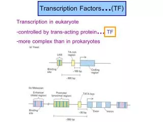
Background • Systems Biology • Understand Biological Networks • Genomic and proteomic methods, bottom-up genetic network engineering • Need to quantitatively characterize unique-element interactions • Many variables • Transient interactions, low-affinity
Summary of Results • Developed high-throughput micro fluidic platform capable of detecting low-affinity transient binding events using mechanically-induced trapping of molecular interactions (MITOMI) • Mapped binding energy landscapes of four eukaryotic bHLH transcription factors • Predicted in vivo function of two TF • Tested base additivity assumption • Tested whether basic region alone determines specificity of bHLH TFs
Experiments • Binding energy landscapes of 4 bHLH TF • MAX iso A • MAX iso B • Pho4p • Cbf1p • bHLH TF generally bind to 5’-CANNTG-3’ E-box • Mid-low nmolar affinities and koff’s of10^-2s^-1
Results Optimal binding sequence = CACGTG MAX iso A 67nM MAX iso B 73.1nM Pho4p 11.1nM Cbf1p 16.6nM
Results MAX B MAX A • Tested additivity assumption • Energetic and informatic role of a base in a given motif is independent of its neighbors • Predicted only 44% of sequences below 2.5kcal/mol
Results • Pho4p and Cbf1p show distinct functions yet have similar recognition sequences • CACGTGsG s=G,C and rTCACGTG r=A,G • Measured possible permutations of 3 flanking base pairs to determine how far base specific recognition extends.
Results • Tested probability of occupancy for regulatory sequence in 5814 yeast genes • Broad agreement with literature
Final Thoughts • Importance of high-throughput methods • Possible limitations of MITOMI
Thank you • Questions?
Fig. S4. (A) Overview of the experimental approach starting with a plain epoxy substrate to be spotted with 2400 spots of a target DNA library. The finished microarray is then aligned and bonded to one of our microfluidic devices after which the surface is prepared, protein synthesized and MITOMI performed. (B) Micrograph of one of the microfluidic unit cells, shown here again for reference. The dashed lines show which regions of the unit cell are schematically depicted in panels C-L. (C) Before any fluid is introduced into the device the chamber valve (green channel in panel B) is closed to prevent flooding of the DNA chamber. (D) Next biotinylated BSA is introduced into our device which covalently bonds to the epoxy functional groups, both activating (via the biotin moieties) and passivating (epoxy groups) the surface. (E) A solution of neutravidin is introduced forming a monolayer on top of the biotinylated BSA layer. (F) The ”button” membrane is closed to protect the detection area from passivation via biotinylated BSA which passivates all accessible surface area. (G) Any unbound biotinylated BSA is purged before the ”button” membrane is opened again allowing access to the neutravidin surface below to which a biotinylated penta-histidine antibody is attached, concluding the surface derivatization. (H) ITT programmed with linear expression template is introduced into the device and allowed to flood the DNA chamber causing the solvation of the stored target DNA. Transcription factor is being synthesized and is pulled down to the surface by penta-histidine antibody. (I) The synthesized transcription factors functionally interact with the solvated target DNA pulling it down to the surface as well. (J) After 60-90 min the ”button” membrane is closed again mechanically trapping any molecular interactions taking place on the surface allowing all solution phase molecules to be washed away without loss of surface bound material (K-L).