Download
1 / 77
850 likes | 1.55k Vues
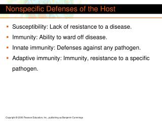
Chapter 21 Nonspecific Body Defenses and Immunity. G.R. Pitts, J.R. Schiller, and James F. Thompson, Ph.D. Defense Systems. Innate (nonspecific) defenses External body membranes Inflammation Antimicrobial proteins, phagocytes and other cells Adaptive (specific) defenses

E N D
Chapter 21Nonspecific Body Defenses and Immunity G.R. Pitts, J.R. Schiller, and James F. Thompson, Ph.D.
Defense Systems • Innate (nonspecific) defenses • External body membranes • Inflammation • Antimicrobial proteins, phagocytes and other cells • Adaptive (specific) defenses • T cells and B cells
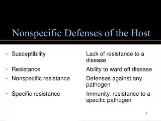
Innate Defense System • Surface Barriers • First line of defense: mechanical and chemical protection • Skin • Mucosal Membranes • Internal Nonspecific Defenses • Second line of defenses • Phagocytes • Natural Killer cells (NK lymphocytes) • Inflammation • Antimicrobial proteins • Fever
Skin and Mucosal Membranes Mechanical Protection • Epidermis • nose hairs, nails • Mucous membranes - line certain organ systems • mucus prevents drying, traps foreign things • respiratory tract cilia sweep mucus out • Lacrimal apparatus -- tear glands and ducts • wash the eye to dilute microbial growth • Saliva - dilute microbes on the oral cavity • Urine - flow dilutes, and acid pH helps kill, microorganisms • Defecation and vomiting - expel toxins and microbes
Skin and Mucosal Membranes Chemical Protection: reduce bacterial growth • Skin • sebum (unsaturated FA’s) forms oily layer • perspiration has fatty acids, salts (NaCl), and mildly acid pH • Lysozyme • in perspiration, tears, saliva, nasal secretions, other tissue fluids • enzyme breaks down bacterial cell walls • Hyaluronic acid • gel-like matrix in most connective tissues • slows the spread of many infectious agents • Gastric juice - stomach nearly sterile due to acid pH, ~2 • Vaginal secretions – mildly acid pH
Innate Defense: Phagocytes • Macrophages (derived from monocytes) are the chief tissue phagocytic cells • Free macrophages wander through tissues in search of microbes and cellular debris • Fixed macrophages: Kupffer cells (liver), microglia (brain), dust cells (lungs) • Neutrophils become phagocytic when encountering infectious material • Eosinophils are weakly phagocytic, deploy destructive granules against parasitic worms
Mechanism of Phagocytosis • Chemotaxis • Adherence – recognition of external carbohydrates and proteins • Aided by opsonins • Ingestion • Killing and digestion
Innate Defense: Natural Killer Cells • Distinct group of large granular lymphocytes (NK lymphocytes = Null Killer lymphocytes) • Nonspecific killers respond to the lack of self-antigens and to the presence of certain surface oligosaccharides • Kill virus-infected body cells and some tumor cells by releasing various defensive molecules – not by phagocytosis • Act before the antigen-specific immune system is activated • Secrete potent chemical signals that enhance the inflammatory response
Innate Defense: Inflammation • Inflammation • Signs: • Redness • Heat • Swelling • Pain • Loss of Function • Function: • Prevent spread of damage • Dispose of pathogens and debris • Set stage for tissue repair
Inflammation Stage 1: Vasodilation and increased vessel permeability • Macrophages and cells lining the gastrointestinal and respiratory tracts carry Toll-Like Receptors (TLRs) that recognize specific classes of microbes • TLReceptor activation causes cytokine release • promotes inflammation & chemotaxis • Mast cells secrete histamine • Other cells secrete various regulatory factors • Histamine, kinins, prostaglandins, leukotrienes, complement • Cause local vasodilation • Increase capillary permeability resulting in edema http://www.komabiotech.co.kr/technical/review/toll_like_receptor.gif
Inflammation: Stage 1 • Edema – increased plasma filtrate seeps into tissue spaces bringing some immune proteins • Helps to dilute harmful substances • Increases supply of oxygen and nutrients needed for metabolism, inflammation and repair • Allows entry of clotting proteins, which reduces the spread of mibrobes
Inflammation Stage 2. Phagocyte moblization • Leukocytosis-inducing factors: increase neutrophil production • Margination (pavementing) • Diapedesis (amoeboid movement) • Chemotaxis of WBCs • neutrophils – rapid arrival • monocytes – slower arrival
Inflammation Stage 3. Tissue repair • Tissue regrowth and repair of damage or scar formation • Pus • dead phagocytes and other WBCs, damaged tissue, and perhaps microbes • if too numerous for effective removal by phagocytes, an abscess may develop
Effects of Inflammation • Increased blood flow results in increased local temperature and local cellular metabolism • Increased capillary permeability and phagocytic migration to the injured tissue
Innate Defense: Antimicrobial Proteins • Attack microorganisms directly • Interfere with microbial reproduction • The most important are: • Interferons • The Complement System • Transferrins which bind Fe2+ in plasma, inhibiting bacterial growth
Interferons (IFNs) • Produced by most tissue cells when infected by a virus • Diffuses to uninfected cells and binds to surface receptors • stimulates macrophages and natural killer lymphocytes • stimulates production of antiviral proteins which block viral replication • inhibits growth of virally infected cells • suppresses growth of tumor cells • Alpha IFN is used against: • hepatitis C virus • herpes virus (genital warts)
The Complement System • 20 plasma and cell membrane proteins that exist as inactive precursors • When activated, the complement system functions to “complement” or enhance certain immune, inflammatory, and allergic responses • Kills bacteria and certain other microbial cell types (our cells normally are protected from complement attack) • Stimulates chemotaxis in leuckocytes • Enhances the effectiveness of both nonspecific and specific defenses
Complement Pathways Classical Pathway is triggered by the specific immune system • Requires binding of antibodies to antigens of invading organisms • Complement C1 then binds to the antigen-antibody complexes (complement fixation) Alternative Pathway is triggered by non-specific interaction among factors B, D, and P, and microbial cell wall polysaccharides (complement fixation) Both pathways involve an enzyme cascade
Complement Pathways • Both pathways converge on C3, which cleaves into C3a and C3b • C3b initiates formation of a membrane attack complex (MAC) • MAC causes cell lysis by creating many hundreds of microscopic holes in the cell’s plasmalemma • C3b is also an opsonin
Innate Defense: Fever • Pyrogens reset the temperature set-point in the hypothalamus • Inhibits some microbes from growing • Increases body’s metabolic rate, which speeds up immune defenses and tissue repair • Increases effects of antimicrobial substances produced by the immune system • Stimulates liver and spleen to sequester iron and zinc (needed by microorganisms) • High fevers are dangerous
Innate Defense System: Review • Surface Barriers • Skin • Mucosal membranes • Internal Nonspecific Defenses • Phagocytes • Natural Killer cells (NK lymphocytes) • Inflammation • Antimicrobial proteins • Fever
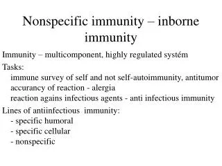
Adaptive Defense • The adaptive immune system: • Acts to immobilize, neutralize, or destroy foreign substances and cells • Amplifies the inflammatory response and activates complement • Is antigen-specific*, systemic, and has memory • *Recognizes specific foreign molecules • Has two interdependent arms • Humoral, or antibody-mediated immunity (AMI) • Cellular, or cell-mediated immunity (CMI)
Adaptive Defense • Definitions: • Immunity: the ability of the body to defend itself against specific foreign invaders (molecules or cells) • Immunogenicity: the ability to stimulate proliferation of specific lymphocytes and specific antibody production • Reactivity: the ability of activated lymphocytes and their products, antibodies, etc., to interact with specific antigens
Adaptive Defense • Definitions: • Specificity: the antigen triggers focused immune defenses (from particular lymphocytes lineages) that respond only to the antigens of this foreign substance/cell • Memory: the immune system produces clones of specific memory lymphocytes (T & B) which react rapidly when the particular foreign substance/cell is encountered again • Specificity and memory differentiate this system from the nonspecific (innate) defenses
Adaptive Defense • Antigen – any substance which provokes specific immune responses • Antigenic determinants • Parts of antigens that trigger the specific immune response • An antigen may be an entire microorganism or only small structures or subregions of large molecules Most “antigens” are complex and express multiple types of antigenic determinants.
Chemical Nature of Antigens • “Complete” Ag: complex macromolecules - usually proteins (nucleo-, lipo-, glyco-) -- sometimes carbohydrates or lipids • Are immunogenic & reactive • “Incomplete” Ag: smaller molecules (haptens) • react with antibodies but cannot cause an immune response without aid (protein carrier) • e.g., poison ivy, drug allergies
Adaptive Defense • Antigen receptor diversity • >1 billion different antigenic determinants are recognized by the body • Genetic recombination shuffles and reorganizes different Ab genes • Major histocompatibility complex antigens (MHC) • unique to each individual’s cells; help in identifying what is self versus foreign • 2 classes of MHC antigens (“markers”) • class I MHC – found on all body cells except RBC's • class II MHC - only on antigen presenting cells (APC’s), thymus cells, and activated T cells
Antigen-Presenting Cells (APCs) • APCs phagocytize, process, and present antigens to lymphocytes • APCs do not respond to specific antigens • APCs contribute to coordinating specific immunity • Macrophages • Dendritic (Langerhans) cells • B lymphocytes The major initiators of adaptive immunity are APCs, which actively migrate to the lymph nodes and secondary lymphoid organs and present antigens to T and B cells
Class I MHC Proteins • Found on all cells, except RBCs • Recognized by Tlymphocytes and APCs • Display peptides from endogenous antigens • Endogenous antigens are: • Associated with body cells • Degraded by proteases and enter the endoplasmic reticulum • Transported through special membrane channels • Bound with MHC class I molecules on the ER membrane • Migrate to the cell membrane as a complex: Ag -- MHC class I molecule
MHC Class I Proteins This is a form of Antigen Presentation Cancer cells often do something quite similar to the virus-infected cells. (Foreign MHC Class I Ags are the source of tissue transplant rejections.)
MHC Class II Proteins • Immune cell identity markers found only on mature B cells, some T cell classes, and antigen-presenting cells • MHC Class II proteins are synthesized in the ER • A phagosome containing a pathogen (with exogenous antigens) merges with a lysosome • MHC Class II proteins migrate into the phagosome where the antigen macromolecules are degraded and particular antigen peptides are bound to the MHC Class II markers • Ag-- MHC class II complex then migrates to the cell membrane and displays antigenic peptide for recognition by CD4 TH cells
MHC Class II Proteins This is a key function of our APCs in most Ag-specific defenses.
Lymphocytes Provide Ag Specificity • B and T lymphocytes develop in bone marrow • Lymphocytes mature and develop immunocompetence (ability to recognize specific antigen) in different locations • B cells mature in the bone marrow and provide Ab-mediated immunity • T cells mature in the thymus and provide cell-mediated immunity
Immunocompetent B or T cells • Naive cells display a unique surface receptor for a specific antigen once mature • Receptor expression occurs before a cell encounters the foreign antigen it may later attack • It is genes, not antigens, that determine which foreign substances our immune system will recognize and resist • Naive cells circulate to secondary lymphoid tissue where they may encounter antigens later • B and T cells become fully functional only after binding with their recognized antigen
Survive Apoptosis Apoptosis Immunocompetent T Cells • T cells mature in the thymus under positive and negative selection pressures • Positive selection – outer thymic cortex • Selects functional T cells which become both immunocompetent and potentially self-tolerant • Non-selected cells die via apoptosis • Negative selection – inner thymic cortex • Kill or regulate off T cells that react with self-antigens
Immunocompetent B Cells • B cells become immunocompetent and self-tolerant in bone marrow • Some self-reactive B cells are killed by apoptosis (clonal deletion) • Some self-reactive B cells can modify their anti-self properties (receptor editing) • Some self-reactive B cells are released from the bone and are inactivated by negative regulation (anergy)
Cell-Mediated Immunity • CMI is involved in most aspects of specific immune defense • Three populations of T lymphocytes regulate specific immunity • Helper TH cells which carry CD4+ markers • Suppressor TS cells • Memory T cells • cytotoxic TC cells which carry CD8+ markers destroy tumor cells and virus-infected cells; they also attack transplanted cells and tissues
Cell -Mediated Immunity Basic steps • Recognition by T lymphocytes of antigen presented by an antigen-presenting cell with matching MHC Class II markers • Proliferation and differentiation of T cells once activated • Production of clones ofidentical effector T cells capable of recognizing a specific antigen • Appropriate action (help, attack, memory, suppression) from T cell subclones
T Cell Activation- Step 1:Antigen Bindingand AntigenPresentation
T Cell Activation- Step 2: Co-Stimulation • T cells must bind to MHC Class II surface receptors on an APC • After co-stimulation with cytokines, T cells enlarge, proliferate, and form clones • Activated T cells differentiate and perform functions according to their T cell class
T Lymphocyte Activity • Primary T cell response usually peaks within a week • T cells then undergo apoptosis within a month • Reduced activity parallels elimination of antigen • This is a negative feedback control • A few Memory T cells remain to respond to any future exposure to the same antigen
Helper TH Lymphocytes • Regulatory cells that play a central management role in the immune response • Once primed by APC antigen presentation, TH cells: • Stimulate proliferation of other T cell classes • Stimulate B cells that have already become bound to antigen • There is NO coordinated immune response without TH cell function
Helper TH Lymphocytes • TH cells interact directly with B cells that have antigen fragments on their surfaces bound to MHC Class II receptors • TH cells express CD4+ cell identity markers • TH cells stimulate B cells to divide more rapidly and begin antibody formation • B cells may be activated without TH cell help by binding to T cell–independent antigens (certain microbial polysaccharides) • Most antigens, however, require TH co-stimulation to activate B cells • Cytokines released by TH amplify nonspecific defenses
Cytotoxic Tc Lymphocytes • TC cells express CD8+ cell identity markers • TC cells, or killer T cells, are the only T cells that can directly attack and kill other cells • They circulate throughout the body in search of body cells that display the antigen to which they have been sensitized • Their targets include: • Virus-infected cells • Cells with intracellular bacteria or parasites • Cancer cells • Foreign cells from blood transfusions (WBCs and platelets) or tissue and organ transplants
Cytotoxic Tc Lymphocytes • Bind to self/anti-self complexes on any body cell • Infected or abnormal cells can be destroyed as long as appropriate antigen and co-stimulatory regulators (e.g., IL-2) are present • [In contrast, Natural Killer cells activate their killing machinery when they bind to a different MHC-related cell surface marker on cancer cells, virus-infected cells, and transplanted cells]
Cytotoxic Tc Lymphocyte Actions • Secrete perforins which cause cell lysis by creating transmembrane pores • Secrete lymphotoxin which fragments the target cell’s DNA • Secrete gamma interferon which stimulates macrophage attack
Suppressor Ts Lymphocytes • TS cells – immune regulatory cells which release cytokines that suppress the activity of both T cells and B cells • Generated when other specific T cell clones are generated • Negative feedback control to bring the body back to normal after the “battle” has been won
Antibody-Mediated Immunity • Antigen challenge – the first encounter between an antigen and a naive B lymphocyte • Antigen presentation usually occurs in the spleen or a lymph node, but can occur in any lymphoid tissue • Antigen presentation usually made by a macrophage, but some B cells can react directly against certain bacterial antigens • Binding of the antigen to the B cell’s specific Ag receptor activates the B cell
Primary Response Activated B cells grow and divide, forming clones bearing the same antigen-specific receptors and secreting the same antigen-specific Ab • Most clone cells become plasma cells that secrete specific antibodies • Clones that do not become plasma cells become B memory cells that can respond to subsequent exposures to the same antigen
Primary Response • Initial B cell differentiation, proliferation, and Ab synthesis requires time after the first Ag exposure • Lag period: 3 to 6 days after antigen challenge • Peak plasma levels of antibody are achieved in ~10 days • Antibody molecules also reach the interstitial fluids, especially where inflammation exists • Antibody levels then decline gradually if there is no additional Ag exposure