Download
1 / 24
240 likes | 365 Vues
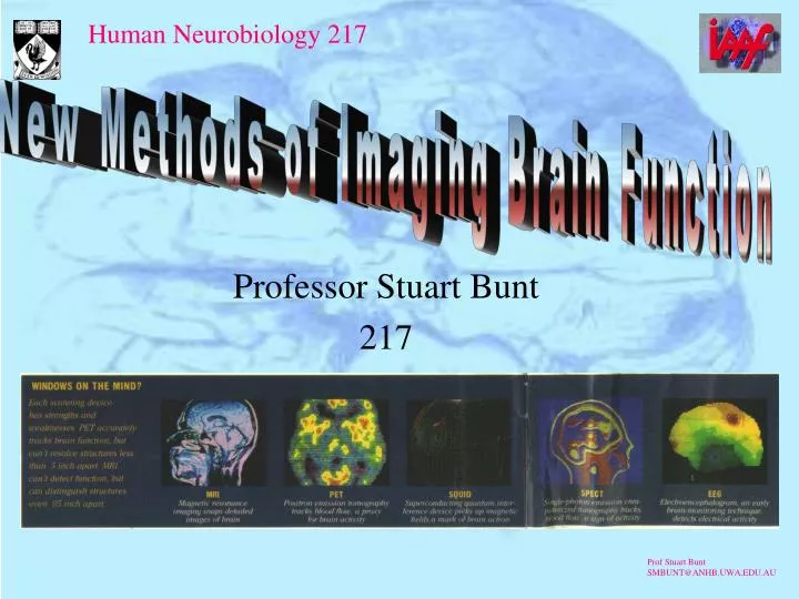
New Methods of Imaging Brain Function. Professor Stuart Bunt 217. Traditional Anatomy. Phrenology, the study of bumps on the skull. Measuring brain weights and size (still being done..see the fuss about Einstein’s brain). Little link between morphology and performance

E N D
New Methods of Imaging Brain Function Professor Stuart Bunt 217
Traditional Anatomy • Phrenology, the study of bumps on the skull. • Measuring brain weights and size (still being done..see the fuss about Einstein’s brain). • Little link between morphology and performance • Neanderthal > Homo sapiens • Hydrocephalic genius
Plane Film X-rays • Not very good for showing soft tissues • Can show bone erosion • Large space filling masses may be picked up if they have largely differing X-ray absorption • Bone of skull requires high energy for penetration
Contrast Media CAT with media around cord Myelogram
Ventriculogram • Air is introduced into the ventricles via the lumbar cistern (in adults) • Via the fontanelles in infants • Patient placed on a tilting table, air fills ventricles • Can detect “space filling” lesions. • Causes severe headaches, same dangers as lumbar puncture.
Subtraction Angiography • Radio opaque medium is introduced into the internal carotid artery or systemically. • Iodine compounds can promote allergic reactions, any arterial invasion dangerous. • Subtract X-ray before contrast medium from X-ray with contrast medium. • Can pick up any abnormality of the blood vessels.
Direct Stimulation • During operations surgeons may have to delimit crucial areas. • Epilepsy often spreads from a temporal lobe focus • The speech area on the planum temporale must be identified. • Cortex stimulated in conscious patient
Computerised Axial Tomography • Many X-rays are taken around the head (or body) • Matching detectors pick up absorbed signal • Computer reconstructs a “slice” • Axial slices can be reconstructed in to a 3D pictures
Magnetic Resonance Imaging 1 • An extremely powerful magnetic field is used to orientate the spin of atoms. • A radio frequency pulse is used to cause some atoms to flip • Energy given off tells a lot about the molecule the atom is in • Can also be used to form images.
CAT vs MRI • MRI (previously known as nuclear magnetic resonance or NMR) is expensive. • The magnet is supercooled with liquid helium in a liquid nitrogen blanket. • Exposures are slow • No metal may enter the field (no pacemakers etc.)
Ultrasound • Ultrasound produced by a piezoelectric crystal or array • No harm? Local heating and cavitation? • Doppler effect raises frequency as blood flows towards detector, lowers frequency in the opposite direction • Can map blood flow speed • Can detect vasospasm (a potentially lethal sequalae to a stroke)
Positron Emission Tomography • Inject a very short lived (radioactive) isotope with extra proton into the internal carotid. • Produced in an (adjacent) cyclotron • Gives out a positron, leaving a neutron. • Positron anhilated when it hits and electron. • Produces two photons in opposite directions • Simultaneity detectors record the event. Choice of labelled compound determines the result (transmitter etc.)
PET Scanner Cyclotron Detector
fMRI • By targeting protons in hydroxyl groups can measure blood flow • Areas of high flow = areas of high activity. • Usual advantages of MRI (see previous slide) • No radioactivity needed, replacing PET
Deep Infrared • New technique • Involves use of far infrared to penetrate the skull • Reflected light picked up • Reflected better by areas of high blood flow • Experimental, low resolution as yet.
SQUIDS • Supercooled, quantum interference devices • Can pick up minute magnetic fields created by the activity of cells. • More localised than EEGs, not an “average” • Low resolution as the detectors are large
EEGs • Extra cerebral recordings average all the electrical activity going on in the brain • Widespread changes can be picked up (epilepsy, coma, REM sleep etc.)
That's All Folks See you in 325? Advanced Neurocience