Download
1 / 38
610 likes | 2.65k Vues
1- EXTRA PYRAMIDAL SYSTEM 2- MOTOR NEURON LESIONS. Lecture 7 DR ZAHOOR ALI SHAIKH. EXTRA PYRAMIDAL SYSTEM. DEFINATION Tracts other than corticospinal tracts are known as EXTRA PYRAMIDAL TRACTS. COMPONENTS OF EXTRAPYRAMIDAL SYSTEM. BASAL GANGLIA BRAINSTEM Giving rise to following tracts:

E N D
1- EXTRA PYRAMIDAL SYSTEM2- MOTOR NEURON LESIONS Lecture 7 DR ZAHOOR ALI SHAIKH
EXTRA PYRAMIDAL SYSTEM DEFINATION • Tracts other than corticospinal tracts are known as EXTRA PYRAMIDAL TRACTS.
COMPONENTS OF EXTRAPYRAMIDAL SYSTEM • BASAL GANGLIA • BRAINSTEM Giving rise to following tracts: • Rubrospinal tract • Vestibulospinal tract • Reticulospinal tract • Tectospinal tract
RUBROSPINAL TRACT • Origin – Red nucleus mid brain • Input - Red nucleus gets input from both cerebellum and cerebral cortical motor areas • Output - Via Rubrospinal tract is directed to contralateral spinal motor neurons ( crosses to opposite side at the level of nucleus and axons are located in lateral spinal white matter anterior to corticospinal tract. • Functions - Involved in movements of distal limbs (hand & feet) also regulates tone and posture. • It is excitatory to flexors and inhibitory to extensor muscles.
VESTIBULOSPINAL TRACT • Location - Vestibular nuclei are located in Pons and Medulla • Input - They receive input from Vestibular apparatus in the inner ear and Cerebellum • Output – Mainly From Lateral vestibular nuclei to spinal cord in Vestibulospinal tract. It remains ipsilatterall • Function - Excitatory to ipsilateral extensor. Inhibitory to flexors muscles • Regulates muscle tone for maintaining balance in response to head movement
RETICULOSPINAL TRACT • Location - Reticular formation in the central grey matter of brain stem • Input - Afferent input to reticular formation comes from spinal cord, vestibular nuclei, cerebellum, Sensory motor cortex, globus pallidus & Lat. Hypothalamus • Output - Descending tract arise from nuclei in pons and medulla 1] Pons – Pontine Reticulospinal tract runs ipsilaterally; Function - Excitatory to Axial extensor muscles 2] Medulla – Medullary reticulospinal tract runs ipsilaterally (some cross also) Function - Inhibitory to axial extensor Muscle
TECTOSPINAL TRACT • Loacation – originates in superior colliculus in midbrain • Input – from visual stimuli • Output - Conveys nerve impulses from superior colliculus (midbrain) to contralateral skeletal muscles that move the head and eyes in response to visual stimuli • Function – Involved in control of neck muscle in response to visual stimuli
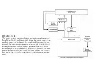
DESCENDING EXTRA PYRAMIDAL MOTOR TRACT TO SPINAL INTERNEURON AND MOTOR NEURON & uncrossed These tracts terminate on anterior horn interneurons. Occasionally they terminate directly on anterior horn motor neurons
EXTRA PYRAMIDAL SYSTEM OR MULTINEURONAL SYSTEM • It has multiple synapses that involve many regions of brain • Final link in extra pyramidal pathway is brain stem, which is influenced by motor cortex, cerebellum, basal nuclei [therefore these brain regions regulate motor activity indirectly] Note – Direct influence on anterior home cell is by primary motor cortex
FUNCTIONS OF EXTRA PYRAMIDAL SYSTEMORMULTINEURONAL SYSTEM • REGULATION OF BODY POSTURE, INVOLVING INVOLUNTARY MOVEMENTS OF LARGE MUSCLE GROUPS OF TRUNK AND LIMBS • REGULATION OF VOLUNTARY MOVEMENT • REGULATION OF TONE
IMPORTANT • Complex and overlapping function exist between Pyramidal and extra pyramidal systems for example while doing fine work like needle work (Pyramidal system) one has to subconsciously assume a particular posture of arms( extra pyramidal system) that enables to do your work
APPLIED • Extra pyramidal tracts some are excitatory and other are inhibitory to muscle tone overall effect – strong inhibitory effect over Gamma Motor Neuron in anterior horn cell What will be the effect of extra pyramidal lesions ? • Hypertonia Why ? - Because strong inhibitory effect over Gamma motor neuron is lost.
Difference between pyramidal and extra pyramidal tracts • EXTRA PYRAMIDAL TRACTS • -Rubrospinal • -Vestibulospinal • -Reticulospinal • -Tectospinal • They originate in Midbrain and brainstem nuclei and have influence of cerbral cortex, basal ganglia and cerebellum which can stimulate or inhibit these nuclei • No direct control of motor cortex or basal ganglia on spinal cord but via nuclei in midbrain and brainstem PYRAMIDAL TRACTS • -Lateral corticospinal -Ant. or ventral corticospinal - Corticobulbar • Cell bodies that contribute to pyramidal tracts are located in precentral gyrus ( Primary, Premotor and supplimentary motor cortex) and somatosensory area. • Pyramidal tract descend directly without synaptic interruption from cerebral motor cortex to spinal cord ( on interneuron and ant. Horn cells)
Difference between pyramidal and extra pyramidal tracts Contd . . . • EXTRA PYRAMIDAL TRACTS • Major extra pyramidal tracts, some cross and others are uncrossed (see table given before) • Function: • Control of body posture involving involuntary movements of axial and Proximal limb muscle PYRAMIDAL TRACTS • 80 % of Corticospinal tracts (lateral) cross in medulla 20 % of corticospinal tract (ventral) cross in spinal cord Because of crossing cerebral cortex controls opposite side of the body • Function: - Lat. Corticospinal tract – fine movement of fingers eg. Writing, needle work - Ventral corticospinal tract – Axial or Postural Movement
COMMON WORDS USED IN NEUROPHYSIOLOGY AND CLINICAL NEUROLOGY • PYRAMIDAL TRACTS • EXTRAPYRAMIDAL TRACTS • PYRAMIDAL LESION – HYPOTONIA Pure pyramidal lesions usually don’t occur in humans • EXTRAPYRAMIDAL LESIONS – HYPERTONIA (Rigidity) • UMN (Upper motor neuron) – Motor tracts coming from Brain to Ant. Horn cells [Pyramidal and extra pyramidal tracts]
Motor System It is two neuron system 1- Upper motor neuron – From motor cortex to anterior horn cell of spinal cord 2- Lower motor neuron – Starts from anterior horn cell and ends on muscle e.g. all peripheral nerves
UMN lesion causes • Increased tone (Spasticity) • Increased reflexes • Clonus: Repetitive contraction and relaxation of muscle in oscillating fashion every second or so • Babinski sign: stimulation of the sole of the foot along outer border causes extension of big toe upward and fanning of other toes (Normally in adults this stimulation causes plantar reflex that is downward flexion of big and small toes.) Note: below one year of age Babinski reflex is normally present. Why ?
Babinski’s sign is hard sign for upper motor neuron lesion, signifies damage to lateral corticospinal tract
LMN • LMN (Lowe motor neuron) – Motor neuron from anterior horn cells to the skeletal muscle( peripheral nerves). • Lower motor neuron also from nuclei of cranial nerves to the skeletal muscles of face and head.
LMN Lesion • LMN lesion (As final pathway to the muscle is damaged) • Decreased tone (Hypotonia / Flaccidity). • Decreased power of the muscles. • Decreased reflexes. • Wasting of muscles.
UMN LESION Paralysis affect movement rather than muscles Muscle wasting is only from disuse, therefore slight. Occasionally marked in chronic severe lesions. Spasticity of clasp-knife’ type. Muscles hypertonic. LMN LESION Individual muscle or group of muscles are affected. Wasting pronounced. Flaccidity. Muscles hypotonic. DIFFERENCE BETWEEN UPPER & LOWER MOTOR NEURON LESION
UMN LESION Tendon reflexes increased. Clonus often present. Superficial reflexes diminished or modified. Abdominal reflex absent. Babinski’s sign +ve, --Increased jaw jerk. LMN LESION Tendon reflexes diminished or absent. Superficial reflexes often unaltered.
COMMON WORDS USED IN NEUROPHYSIOLOGY AND CLINICAL NEUROLOGY • HEMIPLEGIA – Paralysis (loss of power) of half side of the body • HEMIPARESIS – Partial loss of power of half side of the body • PARAPLEGIA – Paralysis in both legs • PARAPARESIS – Partial loss of power in both legs • QUADRIPEGIA – Paralysis in all four limbs • MONOPLEGIA – Paralysis in one limb
R L Lesion of the right dorsal column at L1 produces what impairment? Click for answer Damage to the right dorsal column at L1 causes the absence of light touch, vibration, and position sensation in the right leg. Only fasciculus gracilis exists below T6. Click for explanation
Ipsilateral loss of light touch, vibration, and position sense generalized below the lesion level Below T6 only the fasciculus gracilis is present. Right Dorsal Column Lesion Click to animate DRG R L L1 Dorsal column lesion Common causes include MS, penetrating injuries, and compression from tumors.
R L Lesion of the right lateral spinothalamic tract at L1 produces what impairment? Click for answer Damage to the right lateral spinothalamic tract at L1 causes the absence of pain and temperature sensation in the left leg. Click for explanation
Contralateral loss of pain and temperature sense Right Lateral Spinothalamic Tract Lesion Click to animate DRG R L L1 Lateral spinothalamic tract lesion Common causes include MS, penetrating injuries, and compression from tumors.
R L Lesion of the right lateral corticospinal tract at L1 produces what impairment? Click for answer Damage to the right lateral corticospinal tract at L1 causes upper motor neurons signs (weakness or paralysis, hyperreflexia, and hypertonia) in the right leg. Click for explanation
Ipsilateral upper motor neurons signs generalized below the lesion level UMN signs Weakness (Spastic paralysis) Hyperreflexia (+ Babinski, clonus) Hypertonia Right Lateral Corticospinal Tract Lesion UMN Click to animate R L L1 Lateral corticospinal tract lesion Common causes include penetrating injuries, lateral compression from tumors, and MS.
Complete transection of the right half the spinal cord (Hemicord or Brown-Sequard syndrome) at L1 produces what impairments? R L Click for answer Damage to the right dorsal columns at L1 causes the absence of light touch, vibration, and position sense in the right leg. Damage to the lateral corticospinal tract causes upper motor neuron signs in the right leg (Monoplegia), and damage to the lateral spinothalamic tract causes the absence of pain and temperature sensation in the left leg. Click for explanation
R L Dorsal column lesion Ipsilateral loss of light touch, vibration, and position sense Lateral corticospinal tract lesion Ipsilateral upper motor neurons signs Lateral spinothalamic tract lesion Contralateral loss of pain and temperature sense Hemicord lesion Hemicord Lesion (Brown-Sequard Syndrome) Click to animate L1 Common causes include penetrating injuries, lateral compression from tumors, and MS. Build the lesion
Ipsilateral loss of light touch, vibration, and position sense Ipsilateral upper motor neurons signs Contralateral loss of pain and temperature sense Hemicord lesion Hemicord Lesion (Brown-Sequard Syndrome) UMN Click to animate DRG R L DRG L1 Dorsal column lesion Lateral corticospinal tract lesion Lateral spinothalamic tract lesion